. 2003 Oct;23(19):6958-72.
doi: 10.1128/MCB.23.19.6958-6972.2003.

Repression of the luteinizing hormone receptor gene promoter by cross talk among EAR3/COUP-TFI, Sp1/Sp3, and TFIIB

Affiliations

Repression of the luteinizing hormone receptor gene promoter by cross talk among EAR3/COUP-TFI, Sp1/Sp3, and TFIIB

Ying Zhang et al. Mol Cell Biol. 2003 Oct.

Abstract

Transcription of luteinizing hormone receptor (LHR) gene is activated by Sp1/Sp3 at two Sp1 sites and is repressed by nuclear orphan receptors EAR2 and EAR3 through a direct-repeat (DR) motif. To elucidate the mechanism of the orphan receptor-mediated gene repression, we explored the functional connection between the orphan receptors and Sp1/Sp3 complex, and its impact on the basal transcription machinery. The Sp1(I) site was identified as critical for the repression since its mutation reduced the inhibition by EAR2 and abolished the inhibition by EAR3. Cotransfection analyses in SL2 cells showed that both Sp1 and Sp3 were required for this process since EAR3 displayed a complete Sp1/Sp3-dependent inhibitory effect. Functional cooperation between Sp1 and DR domains was further supported by mutual recruitment of EAR3 and Sp1/Sp3 bound to their cognate sites. Deletion of EAR3 N-terminal and DNA-binding domains that reduced its interaction with Sp1 impaired its inhibitory effect on human LHR (hLHR) gene transcription. Furthermore, we demonstrate interaction of TFIIB with Sp1/Sp3 at the Sp1(I) site besides its association with EAR3 and the TATA-less core promoter region. Such interaction relied on Sp1 site-bound Sp1/Sp3 complex and adaptor protein(s) present in the JAR nuclear extracts. We further demonstrated that EAR3 specifically decreased association of TFIIB to the Sp1(I) site without interfering on its interaction with the hLHR core promoter. The C-terminal region of EAR3, which did not participate in its interaction with Sp1, was required for its inhibitory function and may affect the association of TFIIB with Sp1. Moreover, perturbation of the association of TFIIB with Sp1 by EAR3 was reflected in the reduced recruitment of RNA polymerase II to the promoter. Overexpression of TFIIB counteracted the inhibitory effect of EAR3 and activated hLHR gene transcription in an Sp1 site-dependent manner. These findings therefore indicate that TFIIB is a key component in the regulatory control of EAR3 and Sp1/Sp3 on the initiation complex. Such cross talk among EAR3, TFIIB, and Sp1/Sp3 reveals repression of hLHR gene transcription by nuclear orphan receptors is achieved via perturbation of communication between Sp1/Sp3 at the Sp1-1 site and the basal transcription initiator complex.


Figures

FIG. 1.
FIG. 1.
Alignment of nucleotide sequences of the promoter regions of the hLHR (H) and rLHR (R) genes. DNA sequences of the hLHR and rLHR promoters are shown, and the nucleotides are numbered relative to the translation initiation codon (ATG, +1). The transcriptional start sites are indicated by arrows. The proximal and distal functional Sp1 sites in the human, Sp1(I) and Sp1(II) (in boldface letters) and the corresponding sites in the rat, Sp1-2, and Sp1-4 (boxed letters) are indicated. The DR domains that bind nuclear orphan receptors EAR2, EAR3 (human and rat), and TR4 (human) are underlined by arrows.
FIG. 2.
FIG. 2.
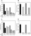
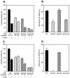
Identification of the Sp1(I) site for EAR2- and EAR3/COUP-TFI-mediated inhibition of the hLHR gene transcription. The wild-type (WT) or Sp1 site mutant hLHR promoter/reporter constructs [Sp1(I)X, Sp1(II)X, and Sp1(I,II)X] was cotransfected with pcDNA3.1 vector or with expression plasmids of EAR2 (A and B) or EAR3/COUP-TFI (C and D) in CV-1 cells. The luciferase activities are expressed as the percentage of the wild-type promoter activity in absence of the orphan receptors (100%, A and C), or as percentage of repression caused by EAR2 or EAR3/COUP-TFI for the individual promoter construct (B and D). The results were normalized by β-galactosidase activity and expressed as the means ± the SE of three independent experiments in triplicate wells.
FIG. 3.
FIG. 3.
Activation of hLHR promoter activity by TR4 independent of the Sp1 site. The wild-type (WT) or Sp1 site mutant hLHR promoter/reporter constructs [Sp1(I)X, Sp1(II)X, and Sp1(I,II)X] was cotransfected with pcDNA3.1 vector only or with expression plasmid of TR4 in CV-1 cells. The luciferase activities are expressed as percentage of the wild-type promoter activity in absence of TR4 (100%), or as percentage of the activation caused by TR4 for the individual construct. The results were normalized by β-galactosidase activity and expressed as the means ± the SE of three independent experiments in triplicate wells.
FIG. 4.
FIG. 4.
Identification of the Sp1(2) site for EAR2-, EAR3/COUP-TFI-mediated repression of the rLHR gene transcription. The wild-type (WT) or Sp1 site mutant rLHR promoter/reporter constructs [Sp1(2)X, Sp1(4)X, and Sp1(2,4)X] was cotransfected with pcDNA3.1 vector only or with expression plasmids of EAR2 (A and B) or EAR3/COUP-TFI (C and D) in CV-1 cells. The luciferase activities are expressed as a percentage of the wild-type promoter activity in the absence of the orphan receptors (100% [A and C]) or as a percentage of the repression caused by EAR2 or EAR3/COUP-TFI for the individual construct (B and D). The results were normalized by β-galactosidase activity and are expressed as means ± the SE of three independent experiments in triplicate wells.
FIG. 5.
FIG. 5.
Requirement of both Sp1 and Sp3 in EAR3/COUP-TFI-mediated repression of the hLHR gene. The wild-type hLHR promoter/reporter gene construct was cotransfected with pACT2 vector only or with increasing dose of EAR3/COUP-TFI expression plasmid in the absence or presence coexpression of Sp1 or Sp3 in Sp1-deficient Drosophila SL-2 cells. The results were normalized as RLU per microgram of protein and are expressed as the means ± the SE of four independent experiments in triplicate wells.
FIG. 6.
FIG. 6.
Analyses of interaction of EAR3 and the Sp1/Sp3 complex in regulation of hLHR gene transcription. (A) In the upper panel are shown DAPAs of mutual recruitment of EAR3 and Sp1/Sp3 complex in the presence of their cognate binding sites (DAPA). JAR nuclear extracts were incubated with 5′ biotin-labeled wild-type Sp1(I) or orphan receptor-binding DR probe or with the corresponding mutant probes (Mutant) devoid of Sp1/Sp3 or EAR3 binding activities. The avidin-precipitated complexes were analyzed by immunodetection with antibodies to Sp1, Sp3, and EAR3. Endogenous expression of the relevant transcription factors was also analyzed by Western blot (W.B.). In the lower panel are shown DNA sequences of the probes utilized in the DAPAs. (B) Schematic diagram of the wild-type (WT) and various deletion constructs of EAR3 (m1, m2, and m3) used in the following Co-IP and functional studies. The numbers represent the amino acid position. Abbreviations: N, N-terminal region; C, C-terminal region; H, hinge region. (C) Co-IP studies of interaction between EAR3 and Sp1. Lysates isolated from JAR cells overexpressing the wild-type (lane WT) or mutant EAR3 constructs (lanes m1, m2, and m3) were used in the Co-IP studies. Monoclonal anti-V5 antibody was utilized to coprecipitate Sp1 protein, followed by immunodetection of Sp1. Normal mouse IgG (lane IgG) was also included as a negative control. (D) Western blot analyses of expression of the wild-type (WT) and mutant EAR3 constructs (m1, m2, and m3) in JAR cells with anti-V5 antibody. (E) Cotransfection studies of the wild-type hLHR gene promoter/reporter gene construct with the wild-type and EAR3 mutant constructs in CV-1 cells. The luciferase activities were expressed as a percentage of the hLHR promoter activity in the absence of EAR3 (100%). The results were normalized by β-galactosidase activity and expressed as the means ± the SE of three independent experiments in triplicate wells.
FIG. 7.
FIG. 7.
Recruitment of basal and trans-transcription factors to the hLHR gene promoter in JAR and CV-1 cells. (A) Recruitment of basal transcription factors, TFIIB and TAFII 250, to the hLHR gene promoter in ChIP assays. Soluble chromatin from JAR cells was precipitated with antibodies against TFIIB, TAFII 250, or preimmune rabbit or mouse immunoglobulin (row IgG). The DNA regions analyzed in PCR are schematically represented in panel E as follows: lanes 1 and 2 refer to 5′ flanking sequences to the promoter; lane 3 covers the full promoter region (positions −176 to +1); lane 4 encompasses the core promoter region only without upstream Sp1 and DR regulatory elements, and a short stretch of coding sequence downstream of ATG (+1); and lane 5 covers solely a part of coding region of the hLHR gene. The results of amplification of soluble chromatin before precipitation were shown as control (Input), and only the negative control with mouse IgG was shown as representative. (B) DAPAs of association of TFIIB or TAFII 250 to the hLHR gene core promoter region. The 5′ biotin-labeled probes utilized encompass DNA sequences of positions −70 to +1 (promoter fragment 1 [PF-1]), positions −70 to −30 (PF-2), and positions −34 to +1 (PF-3), respectively. The probes were incubated with JAR nuclear extracts, followed by immunodetection for bound TFIIB and TAFII 250. The region of the hLHR gene core promoter with arrows indicating the multiple transcription start sites is also shown. (C) ChIP analyses of the recruitment of Sp1, Sp3, EAR3, TFIIB, and RNA Pol II to the hLHR promoter region (region 3 in panel E) in CV-1 and JAR cells. Rabbit IgG was also included as negative control. (D) ChIP analyses of recruitment of Sp1, Sp3, EAR3, and TFIIB to region 6 (in panel E) in CV-1 and JAR cells. Region 6 covers the Sp1 sites and the DR motif of the hLHR gene promoter and some 5′ flanking sequences to the promoter but does not contain the core promoter region. (E) Schematic representation of DNA regions of hLHR gene analyzed in ChIP assays.
FIG. 8.
FIG. 8.
Association of TFIIB with the DR motif and Sp1(I) site of hLHR gene promoter. (A and B) DAPAs were performed to analyze the association of endogenous TFIIB or TAFII 250 to the hLHR gene DR motif (A) or to the Sp1(I) site (B). JAR nuclear extracts were incubated with 5′ biotin-labeled probes, which include the wild-type (WT) and mutant DR elements, and the wild-type and mutant Sp1(I) site. Immunodetection was then carried out with antibodies against TFIIB or TAFII 250. Endogenous expression of TFIIB and TAFII 250 in JAR cells is also shown in Western blot analyses (B [W.B.]). (C) Analyses of protein-protein interaction between TFIIB and Sp1 in DAPAs. Purified Sp1 protein at the indicated doses was incubated with the biotinylated Sp1(I) probe. The excess unbound Sp1 was washed away, and only the DNA-bound form of Sp1 was incubated with 250 ng of purified TFIIB protein. The avidin-precicipated complexes were subject to immunodetection with against TFIIB or Sp1.
FIG. 9.
FIG. 9.
Functional analyses of cross talk among EAR3, Sp1/Sp3, and TFIIB in the regulation of transcription of the hLHR gene. (A) DAPAs were carried out with incubation of JAR nuclear extracts, depleted of TFIIB and EAR3, with 5′ biotin-labeled wild-type Sp1(I) probe, in which a constant amount of 20 ng of recombinant TFIIB protein was added in the presence of 0 to 100 ng of affinity-purified GST/EAR3 fusion protein or GST tag protein only. The avidin-precipitated complexes were subjected to Western blot analyses for immunodetection for TFIIB, Sp1, Sp3, HDAC2, and GST/EAR3. (B) DAPAs of association of TFIIB to the biotinylated PF-1, PF-2, and PF-3 probes (see Fig. 7) in the presence of increasing doses of GST/EAR3 or GST protein. (C) The wild-type (WT) or Sp1 site mutant hLHR promoter constructs [Sp1(I)X, Sp1(II)X, and Sp1(I,II)X] was transfected into JAR cells in absence or presence of pCMV-TFIIB. (D) The wild-type hLHR gene promoter was cotransfected in JAR cells with pCMV-TFIIB, pcDNA3.1-EAR3 expression plasmid, or both. Luciferase activities are expressed as the increase in wild-type promoter activity in the absence of TFIIB and EAR3 (onefold). The results were normalized by β-galactosidase activity and are expressed as the means ± the SE of four independent experiments in triplicate wells.
FIG. 10.
FIG. 10.
Model for the mechanism of Sp1(I) site-dependent silencing of the hLHR gene transcription by EAR3. EAR3 bound to the DR motif interacts with Sp1/Sp3 bound to the Sp1(I) site. Such interaction significantly prevents the robust association of TFIIB to the Sp1(I) site without affecting the recruitment of TFIIB to the hLHR gene core promoter region. Anchoring of the TFIIB at the Sp1(I) site does not require prior binding of EAR3 to its cognate site, since the association is clearly present in the absence of EAR3. We propose that interaction of TFIIB with Sp1/Sp3 is indirectly bridged by a currently unidentified protein(s), as indicated by an open circle (putative tethering protein [PTP]). The EAR3-reduced association of TFIIB to the Sp1/Sp3-DNA complex may induce a nonproductive or less-productive form of PIC, in which the recruitment of RNA Pol II to the hLHR promoter was decreased when the hLHR gene was subjected to a repressed state by EAR3 in JAR cells. Inr, initiator element; TSS, transcriptional start site.

Similar articles

Cited by

References

    1. Bailey, P. J., D. H. Dowhan, K. Franke, L. J. Burke, M. Downes, and G. E. Muscat. 1997. Transcriptional repression by COUP-TF II is dependent on the C-terminal domain and involves the N-CoR variant, RIP13delta1. J. Steroid Biochem. Mol. Biol. 63:165-174. - PubMed
    1. Baniahmad, A., I. Ha, D. Reinberg, S. Tsai, M. J. Tsai, and B. W. O'Malley. 1993. Interaction of human thyroid hormone receptor beta with transcription factor TFIIB may mediate target gene derepression and activation by thyroid hormone. Proc. Natl. Acad. Sci. USA 90:8832-8836. - PMC - PubMed
    1. Black, A. R., J. D. Black, and J. Azizkhan-Clifford. 2001. Sp1 and Kruppel-like factor family of transcription factors in cell growth regulation and cancer. J. Cell Physiol. 188:143-160. - PubMed
    1. Campbell, A. 2002. DNA binding and bending to initiate packaging of phage lambda DNA. Mol. Cell 9:928-929. - PubMed
    1. Catt, K. J., and M. L. Dufau. 1991. Gonadotropic hormones: biosynthesis, secretion, receptors and action, p. 105-155. In S. S. C. Yen and R. B. Jaffe (ed.), Reproductive endocrinology. The W. B. Saunders Co., Philadelphia, Pa.

MeSH terms

LinkOut - more resources