. 2003 Oct;23(19):6973-81.
doi: 10.1128/MCB.23.19.6973-6981.2003.

A phosphomimetic mutation at Ser-138 renders iron regulatory protein 1 sensitive to iron-dependent degradation

Affiliations

A phosphomimetic mutation at Ser-138 renders iron regulatory protein 1 sensitive to iron-dependent degradation

Carine Fillebeen et al. Mol Cell Biol. 2003 Oct.

Abstract

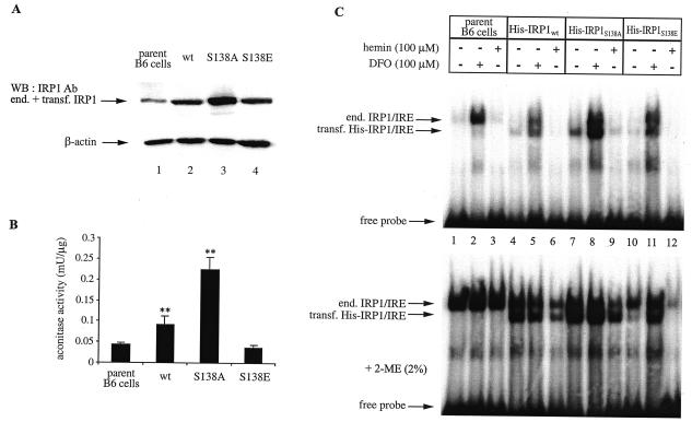
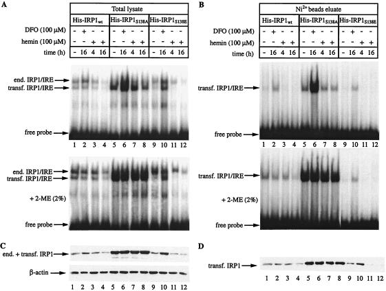
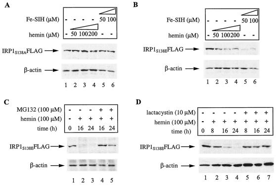
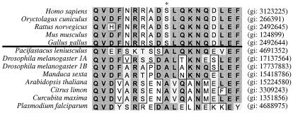
Iron regulatory protein 1 (IRP1) binds to mRNA iron-responsive elements (IREs) and thereby controls the expression of IRE-containing mRNAs. In iron-replete cells, assembly of a cubane [4Fe-4S] cluster inhibits IRE-binding activity and converts IRP1 to a cytosolic aconitase. Earlier experiments with Saccharomyces cerevisiae suggested that phosphomimetic mutations of Ser-138 negatively affect the stability of the cluster (N. M. Brown, S. A. Anderson, D. W. Steffen, T. B. Carpenter, M. C. Kennedy, W. E. Walden, and R. S. Eisenstein, Proc. Natl. Acad. Sci. USA 95:15235-15240, 1998). Along these lines, we show here that a highly purified preparation of recombinant human IRP1 bearing a phosphomimetic S138E substitution (IRP1(S138E)) lacks aconitase activity, which is a hallmark of [4Fe-4S] cluster integrity. Similarly, IRP1(S138E) expressed in mammalian cells fails to function as aconitase. Furthermore, we demonstrate that the impairment of [4Fe-4S] cluster assembly in mammalian cells sensitizes IRP1(S138E) to iron-dependent degradation. This effect can be completely blocked by the iron chelator desferrioxamine or by the proteasome inhibitors MG132 and lactacystin. As expected, the stability of wild-type or phosphorylation-deficient IRP1(S138A) is not affected by iron manipulations. Ser-138 and flanking sequences appear to be highly conserved in the IRP1s of vertebrates, whereas insect IRP1 orthologues and nonvertebrate IRP1-like molecules contain an S138A substitution. Our data suggest that phosphorylation of Ser-138 may provide a basis for an additional mechanism for the control of vertebrate IRP1 activity at the level of protein stability.


Figures

FIG. 1.
FIG. 1.
Recombinant IRP1S138E fails to function as aconitase. (A) SDS-PAGE results of purified human recombinant (rec.) His-tagged wild-type IRP1 (wt), IRP1S138A, and IRP1S138E. The proteins were visualized by staining with Coomassie brilliant blue. (B) Purified recombinant proteins (80 ng) were analyzed by EMSA with a 32P-labeled IRE probe in the absence (top) or presence (bottom) of 2% 2-mercaptoethanol (2-ME). The positions of specific His-IRP1/IRE complexes and excess free probe are indicated by arrows. (C) Aconitase assay prior to (white bars) and after (black bars) treatment with ferrous sulfate-cysteine. Values correspond to results from triplicate samples; the aconitase activity is expressed as milliunits per microgram of recombinant protein. **, P < 0.01 versus values for the control (Student's t test).
FIG. 2.
FIG. 2.
Iron-dependent regulation of chimeric human IRP1 constructs in B6 cells. Wild-type IRP1 (wt), IRP1S138A, and IRP1S138E, tagged with a His6 epitope at their N-termini, were stably transfected (transf.) into B6 cells. (A and B) Cytosolic extracts (devoid of mitochondrial aconitase) of nontransfected parent cells and of cells expressing chimeric wild-type IRP1, IRP1S138A, or IRP1S138E were analyzed by Western blotting (WB) (A) with antibodies (Ab) against IRP1 (top) and β-actin (bottom) and for aconitase activity (B). Values in panel B correspond to results from triplicate samples; the aconitase enzymatic activity is expressed as milliunits per microgram of total protein in the cytosolic lysate. (C) Cells were treated overnight with 100 μM desferrioxamine (DFO) or hemin. Cytoplasmic extracts of nontransfected parent cells (lanes 1 to 3) and of cells expressing chimeric wild-type IRP1 (lanes 4 to 6), IRP1S138A (lanes 7 to 9), or IRP1S138E (lanes 10 to 12) were analyzed by EMSA with a 32P-labeled IRE probe in the absence (top) or presence (bottom) of 2% 2-mercaptoethanol (2-ME). The positions of excess free probe and specific IRP1/IRE complexes, corresponding to endogenous (end.) murine IRP1 and transfected (transf.) human His-IRP1, are indicated by arrows. **, P < 0.01 versus values for the control (Student's t test).
FIG. 3.
FIG. 3.
Hemin-dependent decrease of IRP1S138E expression in B6 cells. Cells expressing human wild-type IRP1, IRP1S138A, or IRP1S138E were treated with 100 μM desferrioxamine (DFO) or hemin for the indicated time intervals. His-tagged chimeric IRP1 was purified from cytoplasmic extracts by affinity chromatography with Ni2+-nitrilotriacetic acid beads. (A and B) Total lysate (A) or affinity-purified chimeric IRP1 (B) was analyzed by EMSA with a 32P-labeled IRE probe in the absence (top) or presence (bottom) of 2% 2-mercaptoethanol (2-ME). The positions of excess free probe and specific IRP1/IRE complexes, corresponding to endogenous (end.) murine IRP1 and transfected (transf.) human His-IRP1, are indicated by arrows. (C and D) Western blotting of total lysates (C) or affinity-purified chimeric IRP1 (D) with antibodies against IRP1 and β-actin (C, bottom).
FIG. 4.
FIG. 4.
Hemin-dependent decrease of IRP1S138E expression in HEK293 cells. The cells were either transiently transfected with pSG5-hIRP1 or stably transfected with pSG5-hIRP1S138A or pSG5-hIRP1S138E to express chimeric wild-type IRP1 (IRP1wt) (A), IRP1S138A (B), or IRP1S138E (C), respectively. All chimeric proteins are tagged with a FLAG epitope at their C termini. The cells were treated with 100 μM hemin for the indicated time intervals, and cell lysates were analyzed by Western blotting (WB) with antibodies (Ab) against IRP1 (top), FLAG (middle), or β-actin (bottom). end., endogenous; transf., transfected.
FIG. 5.
FIG. 5.
(A and B) IRP1S138E, but not IRP1S138A, is sensitive to iron-mediated degradation. HEK293 cells were metabolically labeled for 2 h with [35S]methionine-cysteine and chased with cold media for the indicated time intervals in the absence of iron perturbations (control [ctrl]) or in the presence of 100 μM hemin (+hemin) or 100 μM desferrioxamine (+DFO). The half-life (t1/2) of IRP1S138A (A) or IRP1S138E (B) was assessed by quantitative immunoprecipitation of 500 μg of cytoplasmic extracts with 8.8 μg of FLAG antibody. (C and D) Hemin does not accelerate the turnover of endogenous IRP1 in HEK293 or B6 cells. HEK293 (C) or B6 cells (D) were treated as described for panels A and B, and the decay of endogenous IRP1 was assessed by quantitative immunoprecipitation of 500 μg of cytoplasmic extracts with 20 μl of IRP1 antiserum. Immunoprecipitated material was analyzed by SDS-PAGE on 8% gels, and proteins were visualized by autoradiography. Radioactive bands were quantified by phosphorimaging, and the indicated half-lives were calculated by plotting the data (data not shown).
FIG. 6.
FIG. 6.
Iron-dependent degradation of IRP1S138E is blocked by proteasomal inhibitors. (A and B) HEK293 cells expressing IRP1S138A or IRP1S138E were treated with the indicated concentrations of hemin or Fe-SIH for 16 h. The expression levels of IRP1S138A (A) and IRP1S138E (B) were analyzed by Western blotting with the FLAG antibody (top). (C and D) HEK293 cells expressing IRP1S138E were treated for the indicated time intervals with 100 μM hemin in the presence or absence of 100 μM MG132 (C) or 10 μM lactacystin (D). The expression of IRP1S138E was analyzed by Western blotting with the FLAG antibody (top). All blots shown were also hybridized with an antibody against β-actin (bottom). The cells were viable under all experimental conditions.
FIG. 7.
FIG. 7.
Multiple sequence alignment of amino acids 129 to 147 in IRP1s of various species currently deposited in Swiss-Prot. Ser-138 (asterisk) is conserved only in the IRP1 proteins from vertebrates. The genInfo identifier (gi) numbers of all molecules are indicated on the right. The alignment was made by the Clustal W algorithm (MacVector software, version 6.5.3).

References

    1. Beinert, H., R. H. Holm, and E. Münck. 1997. Iron-sulfur clusters: nature's modular, multipurpose structures. Science 277:653-659. - PubMed
    1. Brazzolotto, X., J. Gaillard, K. Pantopoulos, M. W. Hentze, and J.-M. Moulis. 1999. Human cytoplasmic aconitase (iron regulatory protein 1) is converted into its [3Fe-4S] form by hydrogen peroxide in vitro but is not activated for iron-responsive element binding. J. Biol. Chem. 274:21625-21630. - PubMed
    1. Brown, N. M., S. A. Anderson, D. W. Steffen, T. B. Carpenter, M. C. Kennedy, W. E. Walden, and R. S. Eisenstein. 1998. Novel role of phosphorylation in Fe-S cluster stability revealed by phosphomimetic mutations at Ser-138 of iron regulatory protein 1. Proc. Natl. Acad. Sci. USA 95:15235-15240. - PMC - PubMed
    1. Brown, N. M., M. C. Kennedy, W. E. Antholine, R. S. Eisenstein, and W. E. Walden. 2002. Detection of a [3Fe-4S] cluster intermediate of cytosolic aconitase in yeast expressing iron regulatory protein 1: insights into the mechanism of Fe-S cluster cycling. J. Biol. Chem. 277:7246-7254. - PubMed
    1. Cairo, G., and A. Pietrangelo. 2000. Iron regulatory proteins in pathobiology. Biochem. J. 352:241-250. - PMC - PubMed

Publication types

MeSH terms