. 2003 Sep 30;100(20):11424-8.
doi: 10.1073/pnas.1534703100. Epub 2003 Sep 12.

Mitochondrial positioning in fission yeast is driven by association with dynamic microtubules and mitotic spindle poles

Affiliations

Mitochondrial positioning in fission yeast is driven by association with dynamic microtubules and mitotic spindle poles

Michael P Yaffe et al. Proc Natl Acad Sci U S A. .

Abstract

Microtubules mediate mitochondrial distribution in the yeast Schizosaccharomyces pombe and many higher eukaryotic cells. In higher eukaryotes, kinesin motor proteins have been shown to transport mitochondria along microtubules, but the nature of the mitochondria-microtubule interactions in S. pombe has not been explored. By time lapse, total internal reflection fluorescence microscopy, or spinning-disk confocal microscopy, mitochondria appeared to be both tethered to ends and bound laterally along the sides of microtubules. Mitochondrial tubules extended and retracted when attached to the tips of elongating or shortening microtubules, respectively, but translocation along established microtubules was never observed. Mitochondria that were not associated with microtubules were largely immobile until they were "captured" by a growing microtubule. In mitotic cells, a portion of the mitochondria was tethered to the spindle-pole bodies and moved to the cellular ends during spindle elongation. This association may be important for organelle inheritance during cell division. Thus, in contrast to kinesin-mediated transport used by higher eukaryotes, mitochondrial motility and distribution in fission yeast are driven largely by microtubule polymerization and the elongation of the mitotic spindle.


Figures

Fig. 1.
Fig. 1.
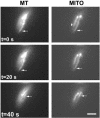
Mitochondria move concomitantly with microtubule polymerization. Cells of S. pombe strain MYP101 induced for expression of GFP-tubulin and RFP targeted to mitochondria were imaged for GFP fluorescence (microtubules) and RFP fluorescence (mitochondria) using TIRF microscopy. (A) Microtubules (MT) and mitochondria (MITO) are shown in successive time-lapse images of a portion of a single cell imaged for 5 s at 20-s intervals. Arrows indicate the growing end of the microtubule and the end of the corresponding mitochondrial tubule. (Bar = 2 μm.) (B–D) Coordinate dynamics of microtubules and mitochondria. The distance of the microtubule tip (open symbols) and the tip of an associated mitochondrial tubule (filled symbols) (relative to an arbitrary position of near the tip of the mitochondrial tubule or the microtubule at time 0) plotted against time. Shown are plots for three distinct microtubule–mitochondrial interactions. In D, the mitochondria are not associated with the microtubule at the beginning and end of the time course.
Fig. 2.
Fig. 2.
Mitochondrial repositioning mediated by microtubule depolymerization. A cell of strain MYP101 was imaged for microtubules (MT) and mitochondria (MITO) as described for Fig. 1. A microtubule undergoing catastrophe and an associated mitochondrion are indicated by the arrows. Also labeled (arrowhead) is a mitochondrial tubule not associated with a microtubule. (Bar = 2 μm.)
Fig. 3.
Fig. 3.
Capture of a mitochondrion by a growing microtubule. Time-lapse images of microtubules (MT) and mitochondria (MITO) were collected as described for Fig. 1, except that they were acquired at 15-s intervals. Arrows indicate a polymerizing microtubule and a portion of a mitochondrial tubule that is repositioned after becoming associated with the microtubule. (Bar = 2 μm.)
Fig. 4.
Fig. 4.
Spindle elongation transports mitochondria toward the cell tips. Cells of S. pombe strain MYP101 induced for expression of GFP-tubulin and RFP targeted to mitochondria were imaged for GFP fluorescence (green; microtubules) and RFP fluorescence (red; mitochondria) by using spinning-disk confocal microscopy. Shown are successive time-lapse images of a mitotic cell captured for 0.5 s at 2.5-min intervals. Arrows indicate one of the cell's spindle pole bodies and its association with mitochondria. Also evident (at times 5.0, 7.5, and 10.0 min) are aster microtubules projecting from spindle-pole bodies, with mitochondria apparently splayed along these transient structures.

Similar articles

Cited by

References

    1. Yaffe, M. P. (1999) Science 283, 1493–1497. - PubMed
    1. Vale, R. D. (1987) Annu. Rev. Cell Biol. 3, 347–378. - PubMed
    1. Nangaku, M., Sato-Yoshitake, R., Okada, Y., Noda, Y., Takemura, R., Yamazaki, H. & Hirokawa, N. (1994) Cell 79, 1209–1220. - PubMed
    1. Yaffe, M. P., Harata, D., Verde, F., Eddison, M., Toda, T. & Nurse, P. (1996) Proc. Natl. Acad. Sci. USA 93, 11664–11668. - PMC - PubMed
    1. Kanbe, T., Kobayashi, I. & Tanaka, K. (1989) J. Cell Sci. 94, 647–656. - PubMed

Publication types

LinkOut - more resources