. 2003 Oct;133(2):813-28.
doi: 10.1104/pp.103.027870. Epub 2003 Sep 4.

Characterization of the targeting signal of the Arabidopsis 22-kD integral peroxisomal membrane protein

Affiliations

Characterization of the targeting signal of the Arabidopsis 22-kD integral peroxisomal membrane protein

Mary A Murphy et al. Plant Physiol. 2003 Oct.

Abstract

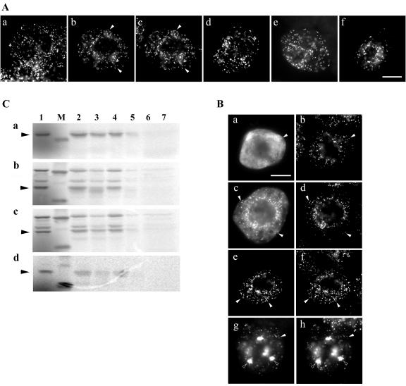
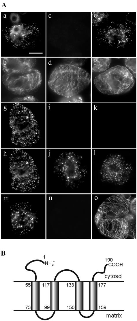
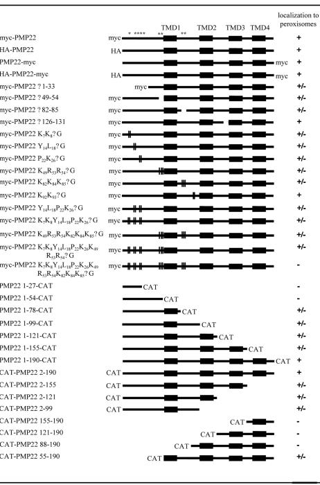
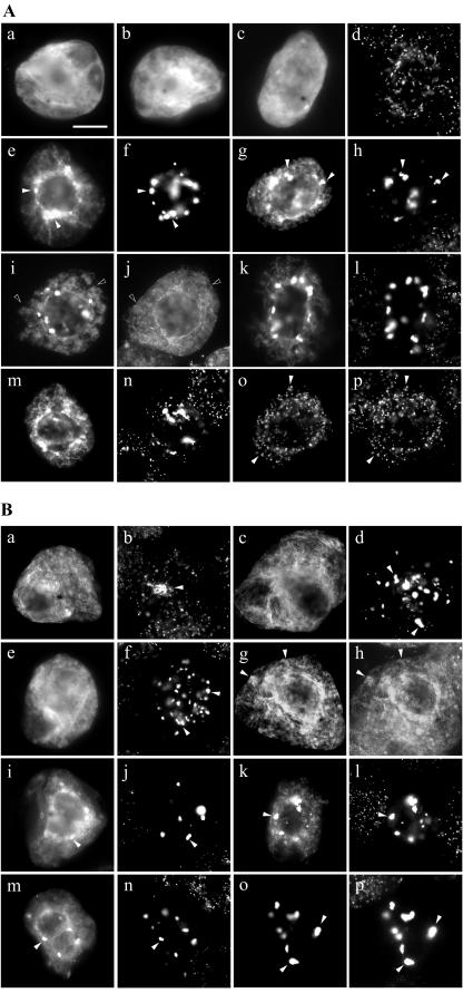
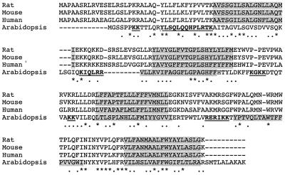
Using a combination of in vivo and in vitro assays, we characterized the sorting pathway and molecular targeting signal for the Arabidopsis 22-kD peroxisome membrane protein (PMP22), an integral component of the membrane of all peroxisomes in the mature plant. We show that nascent PMP22 is sorted directly from the cytosol to peroxisomes and that it is inserted into the peroxisomal boundary membrane with its N- and C-termini facing the cytosol. This direct sorting of PMP22 to peroxisomes contrasts with the indirect sorting reported previously for cottonseed (Gossypium hirsutum) ascorbate peroxidase, an integral PMP that sorts to peroxisomes via a subdomain of the endoplasmic reticulum. Thus, at least two different sorting pathways for PMPs exist in plant cells. At least four distinct regions within the N-terminal one-half of PMP22, including a positively charged domain present in most peroxisomal integral membrane-destined proteins, functions in a cooperative manner in efficient peroxisomal targeting and insertion. In addition, targeting with high fidelity to peroxisomes requires all four membrane-spanning domains in PMP22. Together, these results illustrate that the PMP22 membrane peroxisomal targeting signal is complex and that different elements within the signal may be responsible for mediating unique aspects of PMP22 biogenesis, including maintaining the solubility before membrane insertion, targeting to peroxisomes, and ensuring proper assembly in the peroxisomal boundary membrane.


Figures

Figure 1.
Figure 1.
Peroxisomal targeting and membrane insertion of eptiope-tagged-PMP22s. A, Subcellular localization of endogenous PMP22 and different versions of epitope-tagged Arabidopsis PMP22 in BY-2 cells. Nontransformed (a) or transiently transformed (b-f) BY-2 cells were fixed in formaldehyde, permeabilized with pectolyase and Triton X-100, and incubated in appropriate antibodies. a, Punctate immunofluorescence pattern in nontransformed BY-2 cells incubated with anti-Arabidopsis PMP22 IgGs. b, Transient-expressed myc-PMP22 and endogenous catalase (c) in the same transformed cell; solid arrows indicated obvious colocalizations. Punctate immunofluorescence patterns attributable to expressed HA-PMP22 (d), PMP22-myc (e), and HA-PMP22-myc (f) in transformed cells; colocalizations with endogenous catalase in peroxisomes are not shown. No fluorescence was detected in control experiments including omission of anti-Arabidopsis PMP22, anti-myc, or anti-HA IgGs or mock transformations with pRTL2 vector alone (data not shown). Bar in f = 10 μm. B, Insertion of wild-type and epitope-tagged versions of Arabidopsis PMP22 into isolated peroxisomes in vitro. PMP22 (a), myc-PMP22 (b), HA-PMP22-myc (c), and PMP22-myc (d) were translated in vitro in the presence 35S-Met with the wheat (Triticum aestivum) germ extract system and soluble radiolabeled translation products used in an in vitro import assay with isolated sunflower (Helianthus annuus) peroxisomes. Solid arrows to the left of each panel indicate the location of wild-type or epitope-tagged PMP22 species; the latter was confirmed by immunoprecipitation reactions with anti-myc IgGs (data not shown). Lane 1, Translation products equivalent to 40% of the amount shown in the other lanes. Lane 2, Reisolated radiolabeled protein from an import reaction containing ATP and peroxisomes. Lane 3, The same as lane 2 except that import reactions were treated with the protease thermolysin before reisolation of peroxisomes. Lane 4, Radiolabeled protein reisolated with peroxisomes after an import reaction in the absence of ATP. Lane 5, The same as lane 4 except that import reactions were treated with thermolysin before reisolation of peroxisomes. Lane 6, After an import reaction was carried out in the presence of ATP peroxisomes were reisolated, lysed with the detergent Triton X-100, and treated with thermolysin. Lane 7, The same as lane 5 except that, as a control, peroxisomes were omitted from the mock import reaction. M, Molecular mass markers; upper band (where shown) is 30.1 kD, and the lower band is 20 kD. C, Intracellular sorting of nascent myc-PMP22 from the cytosol to peroxisomes in BY-2 cells. Transiently expressed myc-PMP22 (a, c, e, and g) and endogenous catalase (b, d, f, and h) in transformed cells 5 h (a and b), 12 h (c and d), 20 h (e and f), or 45 h (g and h) after biolistic bombardment. Obvious colocalizations of myc-PMP22 with catalase in individual or globular peroxisomes are indicated with black and white arrows, respectively. Bar in a = 10 μm.
Figure 2.
Figure 2.
Topological orientation of PMP22. A, Immunostaining attributable to transiently expressed myc-PMP22, HA-PMP22-myc, HA-catalase, or to endogenous α-tubulin or catalase in differential permeabilized BY-2 cells. BY-2 cells were formaldehyde fixed, permeabilized with pectolyase and with digitonin (a–d, g–l, n, and o) or Triton X-100 (e, f, and m), and then incubated with appropriate antibodies. Transiently expressed myc-PMP22 (a) and endogenous α-tubulin (b) in the same digitonin-permeabilized transformed cell. Myc-PMP22-bombarded, digitonin-permeabilized cells used for (a and b) immunostained for endogenous peroxisomal matrix catalase (c) and cytosolic α-tubulin (d). Immunostaining of endogenous catalase (e) and α-tubulin (f) in myc-PMP22-bombarded cells permeabilized with Triton X-100. Immunostaining of HA (g) and myc (h) epitopes in a HA-PMP22-myc-transformed cell permeabilized with digitonin. HA-PMP22-myc-bombarded, digitonin-permeabilized cells used for (g and h) immunostained for endogenous catalase (i and k) and the HA (j) or myc epitope (l). Immunostaining of expressed HA-catalase (m) in cells permeabilized with Triton X-100. HA-catalase-bombarded, digitonin-permeabilized cells used for (m) immunostained for the HA epitope (n) and endogenous tubulin (p). Bar in a = 10 μm. B, Predicted topological map of PMP22. Regions of PMP22 proposed to be hydrophobic membrane-spanning domains or hydrophilic domains facing the cytosol or peroxisomal matrix were identified using the TMHMM program (version 2.0) (http://www.cbs.dtu.dk/services/TMHMM-2.0/). Shaded rectangles denote the four membrane-spanning domains (TMD 1–4) and the numbers of their first and last amino acid residues of each TMD are also indicated.
Figure 3.
Figure 3.
Peroxisomal targeting of PMP22 mutant and fusion proteins. Black boxes represent the four predicted TMDs (1–4) in PMP22. Other regions of PMP22 containing putative mPTSs (i.e. amino acids 7 and 8, 14–26, 49–54, and 82–85) are marked with asterisks. Epitope tags and CAT fused to the N or C terminus of PMP22 are indicated. Schematic representations also show deletions or truncations in PMP22 proteins as spaces and site-specific Gly substitutions are marked with vertical bars. The numbers in the name of each fusion construct or myc-PMP22 mutant denotes the specific amino acid residues from PMP22 (1–190 residues) that were fused to the N or C terminus of CAT or delete/replaced with Gly residues. Targeting of PMP22 mutant and fusion proteins to peroxisomes in BY-2 cells was scored as follows: +, exclusively localized to peroxisomes; ±, partially localized to peroxisomes; -, not localized to peroxisomes. Results shown for each construct are a representative of all the transformants (>50) observed from at least two independent biolistic bombardment experiments.
Figure 4.
Figure 4.
Subcellular localization of fusion proteins consisting of different portions of PMP22 fused to the N or C terminus of CAT. BY-2 cells transiently expressing CAT alone, a PMP22-CAT fusion protein, or a CAT-PMP22 (or CAT-APX) fusion protein were formaldehyde fixed, permeabilized with pectolyase and Triton X-100, and processed for immunofluorescence microscopy. A, Subcellular localization of N-terminal PMP22-CAT fusion proteins. Immunostaining attributable to transiently expressed CAT (a), PMP22 1-27-CAT (b), and PMP22 1-54-CAT (c). d, Endogenous peroxisomal catalase staining in the same PMP22 1-54-CAT-transformed cell and neighboring nontransformed cells shown in c. Expressed PMP22 1-78-CAT (e) and CAT-APX (g) and corresponding endogenous catalase (f and h) in transformed cells; black arrows indicate obvious colocalizations. PMP22 1-78-CAT (i) and endogenous ER calreticulin (j) in the same transformed cell; white arrows indicate obvious noncolocalizations. PMP22 1-120 CAT (k), PMP22 1-155 CAT (m), and PMP22 1-190CAT (o) and corresponding endogenous catalase (l, n, and p) in transformed cells; black arrows indicate obvious colocalizations. Bar in a = 10 μm. B, Subcellular localization of C-terminal CAT-PMP22 fusion proteins. Immunostaining attributable to transiently expressed CAT-PMP22 155-190 (a), CAT-PMP22 120-190 (c), CAT-PMP22 88-190 (e), and endogenous peroxisomal catalase (b, d, and f) in the transformed cells; black arrows in b, d, and f denote catalase-containing globular peroxisomes in transformed cells. CAT-PMP22 120-190 (g) and endogenous ER calreticulin (h) in the same transformed cells; black arrows indicated obvious colocalizations CAT-PMP22 2-99 (i), CAT-PMP22 2-121 (k), CAT-PMP22 2-155 (m), CAT-PMP22 2-190 (o), and corresponding endogenous catalase (j, l, n, and p) in transformed cells; black arrows indicate obvious colocalizations.
Figure 5.
Figure 5.
Sequence comparison of PMP22s from rat, mouse, human, and Arabidopsis. Deduced amino acid sequences were obtained from GenBank (accession nos.: rat Q07066; mouse P42925; human AY044439; and Arabidopsis AJ006053) and were aligned using ClustalW and visual inspection. Identical amino acid residues in each of the aligned PMP22s are indicated by asterisks, and similar residues are indicated by dots. TMDs were identified using the TMHMM program (version 2.0). The four predicted TMDs in each of the proteins are shaded and regions tested in Arabidopsis PMP22 in this study to function as mPTSs are in bold and underlined.
Figure 6.
Figure 6.
Subcellular localization of modified versions of myc-PMP22. Transiently transformed BY-2 cells were formaldehyde fixed, permeabilized with pectolyase and Triton X-100, and processed for immunofluorescence microscopy. A, Subcellular localization of myc-PMP22 mutants with alterations in amino acids sequences that resemble positively charged mPTSs in other PMPs. Transiently expressed myc-PMPΔ126-131 (a), myc-PMP22Δ49-54 (c), myc-PMP22Δ82-85 (e), and corresponding endogenous catalase (b, d, and f) in transformed cells; black arrows indicate obvious colocalizations. myc-PMP22Δ49-54 (g) and endogenous ER calreticulin (h) in the same transformed cell; black arrows indicate obvious colocalizations. Coexpressed myc-PMP22Δ49-54 (i) and CAT-APX (j) in the same transformed cell; white arrows indicate obvious noncolocalizations. Myc-PMP22K49R53R54ΔG (k), myc-PMP22K82K84K85ΔG (m), PMP22K49R53R54K82-K84K85ΔG (o), and corresponding endogenous catalase (l, n, and p) in transformed cells; black arrows indicate obvious colocalizations. Bar in a = 10 μm. B, Subcellular localization of myc-PMP22 mutants with Gly substitutions of amino acids sequences that have been proposed to function as mPTSs in mammalian PMP22s. Transiently expressed myc-PMP22K7K8ΔG (a), myc-PMP22K92K93ΔG (c), myc-PMP22Y14L18P22K26ΔG (e), myc-PMP22K7K8Y14L18-P22K26ΔG (g), myc-PMP22Δ1-33 (i), myc-PMP22K7-K8Y14L18P22K26K49K53K54ΔG (k), and corresponding endogenous catalase (b, d, f, h, j, and l) in transformed cells; black arrows indicate obvious colocalizations. Expressed myc-PMP22K7K8Y14L18P22-K26K49K53K54K82K84K85ΔG (m and o) and endogenous catalase (n) or endogenous calreticulin (p) in transformed cells; white arrows in m and n indicate obvious noncolocalization and black arrows in o and p indicate obvious colocalizations.
Figure 7.
Figure 7.
Kinetics of import of various myc-PMP22 mutants in vitro. Selected mutants were tested for their ability to bind to and insert into isolated sunflower peroxisomes. Each mutant myc-PMP22 protein was prepared by in vitro transcription and translation in the presence of 35[S] Met and was incubated with isolated peroxisomes in the presence of ATP at 26°C for the time indicated. For each experiment, two mutant proteins were compared with the parental myc-PMP22 construct to control for any variations in import efficiency between different peroxisome preparations. At the end of the incubation, reactions were returned to ice and treated (+ protease) or not treated (–protease) with thermolysin as described in “Materials and Methods.” After inactivation of the protease, peroxisomes were reisolated through a 0.7 m Suc cushion and were processed for SDS-PAGE and phosphorimaging. “T” translation products equivalent to 40% of the radiolabeled protein added to each of the other incubations. M, Molecular mass markers; the 20-kD marker is shown.

Similar articles

Cited by

References

    1. Baerends RJ, Faber KN, Kram AM, Kiel JAKW, van der Klei IJ, Veenhuis M (2000) A stretch of positively charged amino acids at the N terminus of Hansenula polymorpha Pex3p is involved in incorporation of the protein into the peroxisomal membrane. J Biol Chem 275: 9985–9995 - PubMed
    1. Banjoko A, Trelease RN (1995) Development and application of an in vivo plant peroxisome import system. Plant Physiol 107: 1201–1208 - PMC - PubMed
    1. Brosius U, Dehmel T, Gärtner J (2002) Two different targeting signals direct human peroxisomal membrane protein 22 to peroxisomes. J Biol Chem 277: 774–784 - PubMed
    1. Corpas FJ, Barroso JB, del Rio LA (2001) Peroxisomes as a source of reactive oxygen species and nitric oxide signal molecules in plant cells. Trends Plant Sci 6: 145–150 - PubMed
    1. Coughlan SJ, Hastings C, Winfrey R (1997) Congin and characterization of the calreticulin gene from Ricinus communis L. Plant Mol Biol 34: 897–911 - PubMed

Publication types

LinkOut - more resources