

Laboratory passage and characterization of an isolate of Toxoplasma gondii from an ocular patient in Korea
- PMID: 12972728
- PMCID: PMC2717499
- DOI: 10.3347/kjp.2003.41.3.147
Laboratory passage and characterization of an isolate of Toxoplasma gondii from an ocular patient in Korea
Abstract
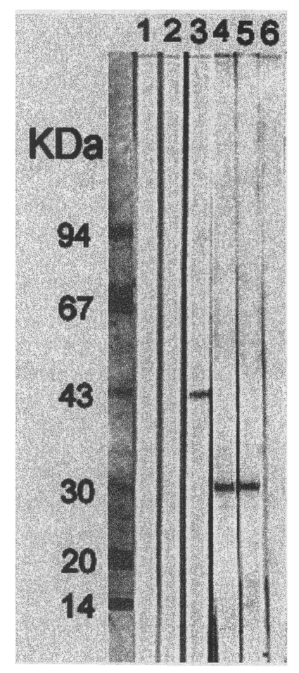
Toxoplasma gondii tachyzoites were isolated from the blood of an ocular patient, and have been successfully passaged in the laboratory, for over a year, by peritoneal inoculation in mice. The isolated parasite was designated the Korean Isolate-1 (KI-1) and its characteristics were compared with those of the RH strain, a wellknown virulent strain originating from a child who suffered from encephalitis. The morphology, pathogenicity, infectivity and cell culture characteristics of the KI-1 were similar to those of the RH strain. Both RH and KI-1 antigens were detected by an anti-T. gondii monoclonal antibody (mAb), Tg563, against the major surface protein SAG1 (30 kDa), whereas no reaction was observed against an anti-Neospora caninum mAb, 12B4. The KI-1 was confirmed as an isolate of T. gondii. A long-term laboratory maintenance and characterization of a local T. gondii isolate is reported for the first time in the Republic of Korea.
Figures


References
-
- Amendoeira MRR, Coutinho SG. Isolation ofToxoplasma gondii from the saliva and tonsils of a three-year-old child. J Infect Dis. 1982;145:587. - PubMed
-
- Chai JY, Kook J, Guk SM, Chang YP, Yun CK. Experimental infection of murine splenic lymphocytes and granulocytes with Toxoplasma gondii RH tachyzoites. Korean J Parasitol. 1997;35:79–85. - PubMed
-
- Choi JS, Choi CS, Soh CT. Isolation of Toxoplasma gondii from congenital and acquired chorioretinitis cases. Yonsei Rep Trop Med. 1980;11:39–42.
-
- Choi WY, Nam HW, Kwak NH, et al. Foodborne outbreaks of human toxoplasmosis. J Infect Dis. 1997;175:1280–1282. - PubMed
-
- Choi WY, Nam HW, Youn JH, et al. Detection of antibodies in serum and cerebrospinal fluid to Toxoplasma gondii by indirect latex agglutination test and enzyme-linked immunosorbent assay. Korean J Parasitol. 1992;30:83–90. - PubMed
Publication types
MeSH terms
Substances
LinkOut - more resources
Full Text Sources
