

doi: 10.1038/nn1120.
Epub 2003 Sep 14.
Developmental acquisition of sensory transduction in hair cells of the mouse inner ear
Affiliations
- PMID: 12973354
- PMCID: PMC2669437
- DOI: 10.1038/nn1120
Item in Clipboard
Developmental acquisition of sensory transduction in hair cells of the mouse inner ear
Nat Neurosci.
2003 Oct.
Abstract
Sensory transduction in hair cells requires assembly of membrane-bound transduction channels, extracellular tip-links and intracellular adaptation motors with sufficient precision to confer nanometer displacement sensitivity. Here we present evidence based on FM1-43 fluorescence, scanning electron microscopy and RT-PCR that these three essential elements are acquired concurrently between embryonic day 16 and 17, several days after the appearance of hair bundles, and that their acquisition coincides with the onset of mechanotransduction.
Figures
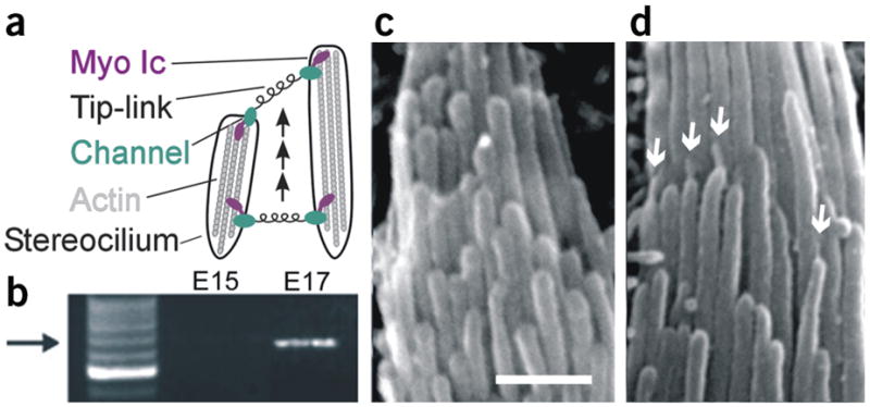
Acquisition of transduction, myosin Ic and tip-links. (a) Diagram illustrating the model for transduction development. (b) Myosin Ic RT-PCR products obtained using mRNA extracted from eight E15 and nine E17 utricles. Arrow indicates position of expected 660-bp product sequenced to confirm identity. (c,d) Scanning electron micrographs of representative hair bundles at E15 and E17, respectively. Scale bar, 1 μm. Arrows indicate several tip-links.
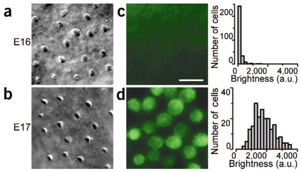
DIC and FM 1-43 fluorescence images and analysis of E16 and E17 hair cells. (a,b) DIC image of E16 and E17 hair bundles viewed from above. (c,d) Fluorescence image of E16 and E17 hair cells focused at the cell body level. Scale bar (10 μm) applies to all images. Right, histograms of the mean fluorescence of 289 E16 (top) and 208 E17 (bottom) cells. Bin width, 200 arbitrary units (a.u.).
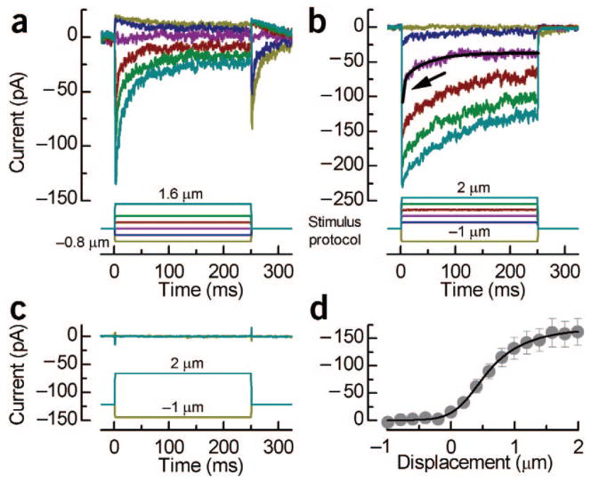
Transduction currents from embryonic hair cells. (a) Family of transduction currents from an E17 hair cell showing the fastest myosin Ic–type adaptation we observed, an open probability of ~10%, adaptation to negative deflections and rebound currents at the end of the step. (b) Transduction currents from an E17 cell showing slow and fast adaptation. The purple trace was fit by a double exponential (black line) with time constants of 2.8 ms (arrow) and 43.6 ms. (c) Representative currents recorded from an E15 hair cell. (d) Mean current–displacement relationship from eight E17 hair cells fit with a second-order Boltzmann equation (10–90% operating range, 1.36 μm; resting open probability, 8.4%). All experiments were approved by the University of Virginia Animal Care and Use Committee.
References
-
- Denman-Johnson K, Forge A. J Neurocytol. 1999;28:821–835. - PubMed
-
- Denk W, Holt JR, Shepherd GM, Corey DP. Neuron. 1995;15:1311–1321. - PubMed
-
- Holt JR, et al. Cell. 2002;108:371–381. - PubMed
-
- Kros CJ, et al. Nat Neurosci. 2002;5:41–47. - PubMed
-
- Sahly I, El-Amraoui A, Abitbol M, Petit C, Dufier JL. Anat Embryol (Berl) 1997;196:159–170. - PubMed
Publication types
MeSH terms
Substances
Grants and funding
LinkOut - more resources
Full Text Sources
