. 2003 Oct;73(4):957-66.
doi: 10.1086/378781. Epub 2003 Sep 12.

Mutations in capillary morphogenesis gene-2 result in the allelic disorders juvenile hyaline fibromatosis and infantile systemic hyalinosis

Affiliations

Mutations in capillary morphogenesis gene-2 result in the allelic disorders juvenile hyaline fibromatosis and infantile systemic hyalinosis

Oonagh Dowling et al. Am J Hum Genet. 2003 Oct.

Abstract

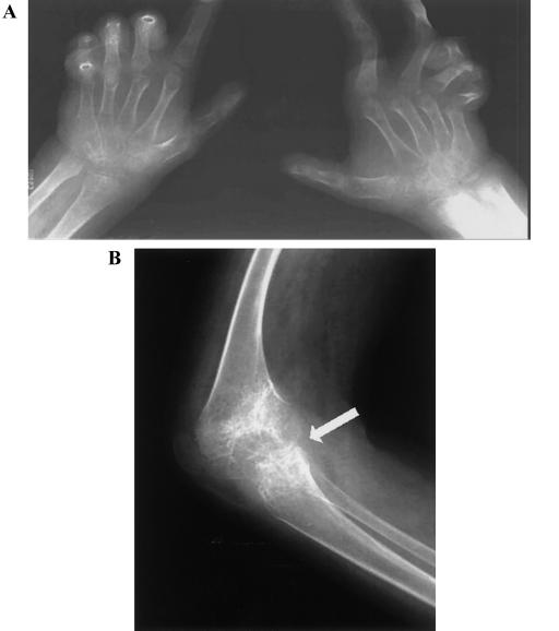
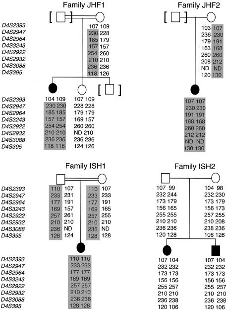
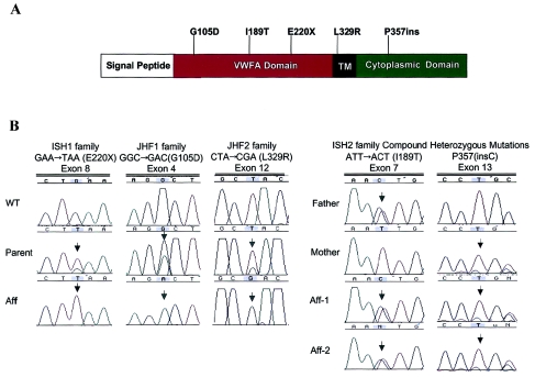
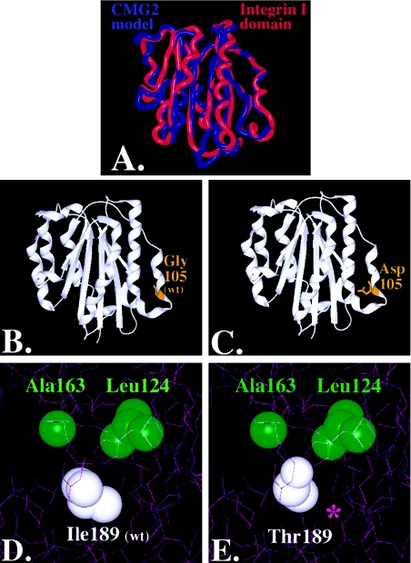
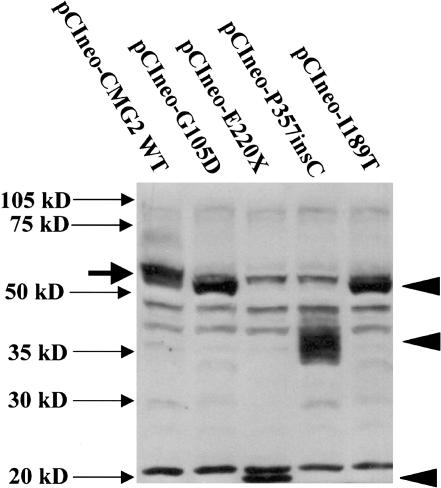
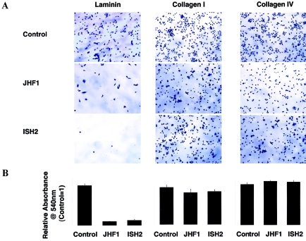
Juvenile hyaline fibromatosis (JHF) and infantile systemic hyalinosis (ISH) are autosomal recessive syndromes of unknown etiology characterized by multiple, recurring subcutaneous tumors, gingival hypertrophy, joint contractures, osteolysis, and osteoporosis. Both are believed to be allelic disorders; ISH is distinguished from JHF by its more severe phenotype, which includes hyaline deposits in multiple organs, recurrent infections, and death within the first 2 years of life. Using the previously reported chromosome 4q21 JHF disease locus as a guide for candidate-gene identification, we identified and characterized JHF and ISH disease-causing mutations in the capillary morphogenesis factor-2 gene (CMG2). Although CMG2 encodes a protein upregulated in endothelial cells during capillary formation and was recently shown to function as an anthrax-toxin receptor, its physiologic role is unclear. Two ISH family-specific truncating mutations, E220X and the 1-bp insertion P357insC that results in translation of an out-of-frame stop codon, were generated by site-directed mutagenesis and were shown to delete the CMG-2 transmembrane and/or cytosolic domains, respectively. An ISH compound mutation, I189T, is predicted to create a novel and destabilizing internal cavity within the protein. The JHF family-specific homoallelic missense mutation G105D destabilizes a von Willebrand factor A extracellular domain alpha-helix, whereas the other mutation, L329R, occurs within the transmembrane domain of the protein. Finally, and possibly providing insight into the pathophysiology of these diseases, analysis of fibroblasts derived from patients with JHF or ISH suggests that CMG2 mutations abrogate normal cell interactions with the extracellular matrix.


Figures

Figure  1
Figure 1
Radiological features of affected individual in family JHF1. A, Frontal view of both hands, revealing diffuse osteopenia and narrowing of interarticular spaces. Multiple subluxations and contractures are present. B, Lateral view of the knee, revealing marked narrowing of the joint space (arrow) and profound osteopenia.
Figure  2
Figure 2
Analysis of pedigrees and haplotypes in four families with JHF or ISH. Genotypes are represented by allele sizes (bp), and markers are shown according to their physical order. Blackened symbols denote affected individuals, and shaded areas denote disease-segregating haplotypes.
Figure  3
Figure 3
A, Predicted CMG-2 protein domains. The protein is 487 amino acids in length and contains an N-terminal signal peptide, followed by a VWFA domain, a TM domain, and a cytosolic tail. Mutations were identified in exons 3, 7, 8, and 12 and are shown relative to affected protein domains. B, DNA sequence analysis of CMG-2 in individuals with ISH and JHF. Three homozygous mutations were identified: GAA→TAA (E220X) nonsense mutation in exon 8 of ISH1 family, GGC→GAC (G105D) missense mutation in exon 4 of JHF1 family, and CTA→CGA (L329R) missense mutation in exon 12 of JHF2 family. Both affected children in family ISH2 were compound heterozygotes: ATT→ACT (I189T) missense mutation (paternal allele) and a nucleotide insertion (P357insC) (maternal allele).
Figure  4
Figure 4
Molecular modeling of CMG-2 mutations: superposition of CMG-2 model (red) with chain A of the Alpha-X Beta2 Integrin I Domain (PDB accession number 1N3Y) (blue). Nonconserved residues were mutated using the software program O (Jones et al. 1991), and the CMG-2 model was energy minimized using Molecular Operating Environment software (A). The root mean square deviation of the CMG-2 model from the integrin template is ∼.103 nm, with greater variation in the loops and less variance in the conserved regions where the mutations reside. Glycine 105 (B) was mutated to an aspartate (C), within the extracellular region and rendered with SPOCK and Raster3D (Merritt and Bacon 1997). Isoleucine 189 (D) was mutated to threonine (E), and contours were provided by the calculated electron density. A cavity is formed, as indicated by the purple asterisk (*) in panel E.
Figure  5
Figure 5
CMG-2 mutations, resulting in altered CMG-2 protein expression, as detected by western blotting. Two hundred ninety-three cells were transfected with 1.5 mg of plasmid DNA (in six-well dishes) with Lipofectamine 2000 and various CMG-2 WT and mutant constructs, as indicated. Following transfection, cells were lysed after 24 h with 0.5 ml SDS-PAGE sample buffer, containing mercaptoethanol, and were treated at 100°C for 10 min. Thirty milliliters of sample was loaded per lane on a 10% SDS-PAGE gel, and protein samples were transferred to PVDF membranes and were probed with anti-CMG-2 affinity-purified antibodies (1 mg/ml), as described elsewhere (Bell et al. 2001). Closed arrowheads indicate the position of anti-CMG-2 reactive mutant proteins. Solid arrow indicates the position of CMG-2 WT protein observed in 293 cells transfected with pCIneo-CMG2-WT.
Figure  6
Figure 6
Crystal violet staining of adherent patient and control primary fibroblasts to laminin, collagen I, and collagen IV ECM. Cells were plated in serum-free media at a density of 1×10⁵ cells/well and were allowed to adhere to laminin, collagen I, and collagen IV 24-well plates (BD Biosciences) for 75 min. Unbound cells were removed by washing with PBS, and adherent cells were fixed in ethanol (10 min), were stained with 0.5% crystal violet (20 min), were washed extensively with water, and were solubilized with 800 μl 1% SDS. Relative adhesion was quantified by monitoring the absorbance of released dye at 540 nm (n=4). Experiments were repeated three times in quadruplicate. Cells are shown at 7.5× magnification. Bar charts indicate adhesion of patient cells compared with control cells.

References

Electronic-Database Information

    1. Celera, http://www.celera.com/ (for identification of candidate genes)
    1. Center for Medical Genetics, Marshfield Medical Research Foundation, http://research.marshfieldclinic.org/genetics/
    1. Decode, http://www.decodegenetics.com/ (for the human genetic map)
    1. Online Mendelian Inheritance in Man (OMIM), http://www.ncbi.nlm.nih.gov/Omim/ (for JHF, ISH, epidermolysis bullosa letalis, epidermolysis bullosa with pyloric atresia, and multiple epiphyseal dysplasia)
    1. Protein Data Bank, http://www.rcsb.org/pdb/

References

    1. Aurora R, Rose GD (1998) Helix capping. Protein Sci 7:21–38 - PMC - PubMed
    1. Bell SE, Mavila A, Salazar R, Bayless KJ, Kanagala S, Maxwell SA, Davis GE (2001) Differential gene expression during capillary morphogenesis in 3D collagen matrices: regulated expression of genes involved in basement membrane matrix assembly, cell cycle progression, cellular differentiation and G-protein signaling. J Cell Sci 114:2755–2773 - PubMed
    1. Chapman KL, Mortier GR, Chapman K, Loughlin J, Grant ME, Briggs MD (2001) Mutations in the region encoding the von Willebrand factor A domain of matrilin-3 are associated with multiple epiphyseal dysplasia. Nat Genet 28:393–396 - PubMed
    1. Colognato H, Yurchenco PD (2000) Form and function: the laminin family of heterotrimers. Dev Dyn 218:213–234 - PubMed
    1. Daluiski A, Engstrand T, Bahamonde ME, Gamer LW, Agius E, Stevenson SL, Cox K, Rosen V, Lyons KM (2001) Bone morphogenetic protein-3 is a negative regulator of bone density. Nat Genet 27:84–88 - PubMed

Publication types

Associated data