. 2003 Aug 21;48(16):2577-89.
doi: 10.1088/0031-9155/48/16/301.

High power transcranial beam steering for ultrasonic brain therapy

Affiliations

High power transcranial beam steering for ultrasonic brain therapy

M Pernot et al. Phys Med Biol. .

Abstract

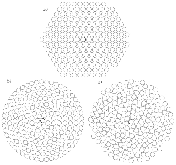
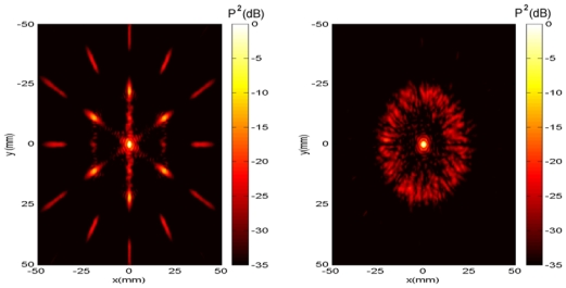
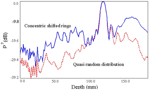
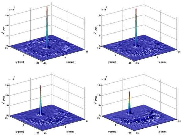
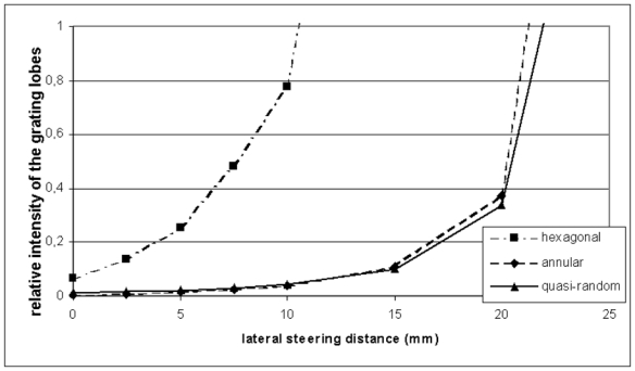
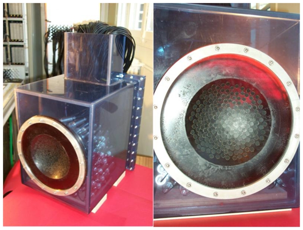
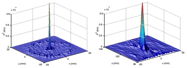
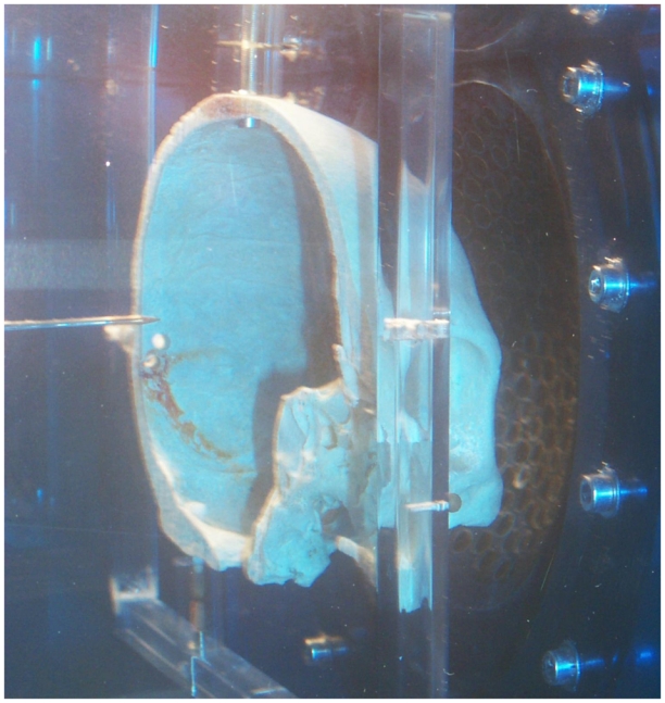
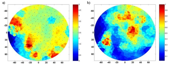
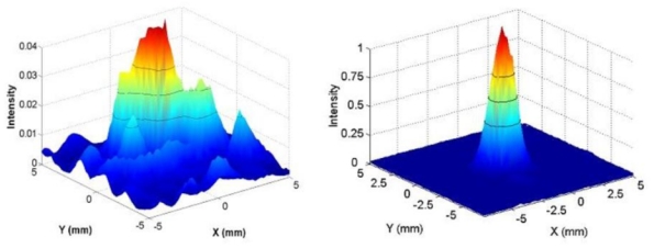
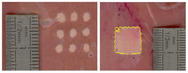
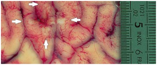
A sparse phased array is specially designed for non-invasive ultrasound transskull brain therapy. The array is made of 200 single elements corresponding to a new generation of high power transducers developed in collaboration with Imasonic (Besançon, France). Each element has a surface of 0.5 cm2 and works at 0.9 MHz central frequency with a maximum 20 W cm(-2) intensity on the transducer surface. In order to optimize the steering capabilities of the array, several transducer distributions on a spherical surface are simulated: hexagonal, annular and quasi-random distributions. Using a quasi-random distribution significantly reduces the grating lobes. Furthermore, the simulations show the capability of the quasi-random array to electronically move the focal spot in the vicinity of the geometrical focus (up to +/- 15 mm). Based on the simulation study, the array is constructed and tested. The skull aberrations are corrected by using a time reversal mirror with amplitude correction achieved thanks to an implantable hydrophone, and a sharp focus is obtained through a human skull. Several lesions are induced in fresh liver and brain samples through human skulls, demonstrating the accuracy and the steering capabilities of the system.


Figures

Fig. 1
Fig. 1
(a) Hexagonal (b) annular and (c) quasi-random transducer distributions.
Fig. 2
Fig. 2
Pressure distribution in the focal plane for the hexagonal (left) and quasi-random (right) arrays. The pressure squared is in dB scale.
Fig. 3
Fig. 3
Maximum pressure squared (in dB scale) reached on the beam axis for the concentric ring and quasi random arrays.
Fig. 4
Fig. 4
Intensity distribution in the focal plane for the quasi-random array, with the focus moved electonically at (a) 0 mm (b) 5mm (c) 10mm and (d) 15 mm in the y lateral direction.
Fig. 5
Fig. 5
Maximum intensity of the grating lobes when the focus is steered from 0 to 20 mm off the beam axis. The values are normalized by the intensity of the main lobe. Solid line: quasi random array. Dotted line: annular array. Dash-dotted line: hexagonal array.
Fig. 6
Fig. 6
The 200-element sparse array prototype and the electronic system. The waterproof PVC cube contains the 200 electrical matching boxes. On the right: the quasi random distribution of transducers embedded in spherical shaped Ureol.
Fig. 7
Fig. 7
(a) Experimental scan of the pressure squared for the quasi-random array in the focal plane (XY plane). This beam pattern was found to be in good agreement with the numerical simulation in Fig. 4. (b) Experimental scan of the pressure squared in the longitudinal XZ plane. The white lines correspond to –3dB and –6dB.
Fig. 8
Fig. 8
Experimental setup for the time reversal process. On the left side: the needle hydrophone (PZT, 0.4 mm, SEA) used for the corrections of the skull aberrations. On the right side: the high power 200-elements ultrasonic array. The whole experiment is performed in degassed water.
Fig. 9
Fig. 9
2D spatial map of the wavefront aberrations induced by the skull. Typically the celerity in the skull can reach 3500 m.s−1 in some locations and the absorption 8 dB.mm−1. The black dots represents the projection of the transducers positions. (a) The phase distortions (μs) (b) The relative amplitude distortion.
Fig. 10
Fig. 10
Experimental distribution of the intensity in the focal plane (a) without aberrations corrections and (b) with corrections. Black lines correspond to -3 dB, -6 dB and -9 dB.
Fig. 11
Fig. 11
Thermally induced lesions in fresh liver through a human skull. The target is located at depth Z = 120 mm from the array. In both experiments the aberration corrections were achieved at center by using an implanted hydrophone and the other impacts were achieved by tilting electronically the beam. a) Lesion size is about 2 mm in diameter for the center spot. The spatial step between each lesion is 5 mm. b) Square-shaped necrosis performed with 25 electronically steered focus (2mm spatial step). The thermal dose has been calculated for this experiment. The contour delimits the zone where the thermal dose is higher than 243 min. It predicts the size of the necrosis in good agreement with the real necrosis.
Fig. 12
Fig. 12
Thermally induced lesions in a sheep brain through a human skull. The target is located at depth Z = 120 mm from the array. The aberration corrections were achieved at center by using an implanted hydrophone and next impacts were achieved by tilting electronically the beam along a cross. On the vertical and the horizontal lines the step between each lesion was 0.2 mm. The white arrows point the extremity of the cross.

Similar articles

Cited by

References

    1. Aubry J-F, Tanter M, Pernot M, Thomas J-L, Fink M. Experimental demonstration of non invasive transskull adaptive focusing based on prior CT scans. J Acoust Soc Am. 2003;113(1):85–93. - PubMed
    1. Basauri L, Lele PP. A simple method for production of trackless focal lesions with focused ultrasound: Statistical evaluation of the effects of irradiation on the central nervous system of the cat. J Physiology. 1962;160:513–514. - PMC - PubMed
    1. Cassereau D, Guyomar D. Computation of the impulse diffraction of any obstacle by impulse ray modelling – Prediction of the signals distortions. J Acoust Soc Am. 1988;84(4):1504–1516.
    1. Chapelon J-Y, Cathignol D, Cain C, Ebbini E, Kluiwstra J, Sapozhnikov OA, Fleury G, Berriet R, Chupin L, Guey J-L. New piezoelectric transducers for therapeutic ultrasound. Ultrasound Med Biol. 2000;26(1):153–159. - PubMed
    1. Clement GT, White JP, Hynynen K. Investigation of a large area phased array for focused ultrasound surgery through the skull. Phys Med Biol. 2000;45:1071–1083. - PubMed

Publication types