. 2003 Oct;134(1):13-22.
doi: 10.1046/j.1365-2249.2003.02254.x.

Delivery of antigen in allogeneic cells preferentially generates CD(4+) Th1 cells

Affiliations

Delivery of antigen in allogeneic cells preferentially generates CD(4+) Th1 cells

J N Agrewala et al. Clin Exp Immunol. 2003 Oct.

Abstract

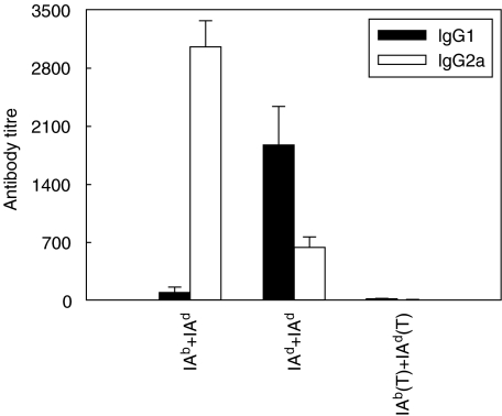
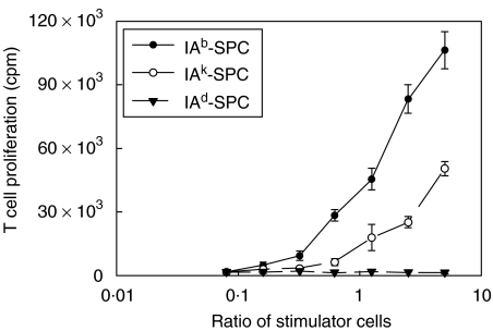
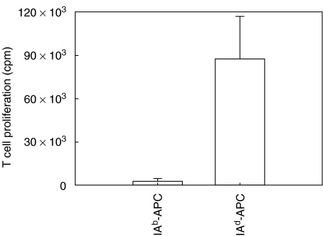
We have examined the possibility of evoking antigen-specific T cell immune response by using allogeneic cells as a source of adjuvant and also as a vehicle to deliver antigen. The mice were immunized with different preparations of antigen-pulsed allogeneic and syngeneic splenocytes. It was observed during the study that the animals immunized with antigen-pulsed mitomycin C treated allogeneic cells elicited antigen specific CD(4+) Th1 cell response. Predominant release of IL-2, interferon (IFN)-gamma and IgG2a-isotype also occurred. In contrast, mice immunized with antigen-pulsed syngeneic cells chiefly enhanced the production of interleukin (IL)-4 and IgG1-isotype. Further, allogeneic macrophages induced better T cell response than B cells or splenocytes and prominently induced the expression of B7-1 and B7-2. Immunization with antigen-pulsed macrophages provided better recall responses compared to B cells. This was manifested by the high LFA-1alpha and low CD45RB expression on T cells. Because it is already known that mitomycin C-treated cells undergo apoptosis and dendritic cells engulf apoptotic cells, we therefore propose that generation of T cell response using antigen-pulsed allogeneic cells may be due to the engulfment of these cells by dendritic cells, which may then process and present antigen entrapped in allogeneic cells to activate naive CD(4+) T cells and differentiate them to Th1 cells. This study therefore provides a rational basis for manipulating antigen-specific responses by immunizing with antigen-pulsed allogeneic cells.


Figures

Fig. 1
Fig. 1
Primary immunization with antigen-pulsed syngeneic cells elicits antigen-specific CD4+ T cell proliferation. Different groups of Balb/c mice received primary immunization with OVA-pulsed (100 µg/ml), mitomycin C treated 1 × 10⁶ allogeneic splenocytes expressing either IAb or IAk haplotype, or syngeneic splenocytes (IAd) (a). The following four experimental groups were utilized for secondary immunization: (i) IAd + IAd, primary and secondary immunizations with antigen-pulsed syngeneic splenocytes; (ii) IAb + IAd, primary and secondary immunizations with antigen-pulsed allogeneic and syngeneic splenocytes, respectively; (iii) IAb + IAk, primary (IAb) and secondary (IAk) immunizations with antigen-pulsed allogeneic splenocytes; (iv) IAd + IAb, primary and secondary immunizations with antigen-pulsed syngeneic and allogeneic splenocytes, respectively (b). In the multiple boosters each group received three inoculations, as described in the inset (c). The animals were sacrificed 1 week after the last injection; CD4+ T cells (5 × 10⁴/well) were isolated, pooled and cultured with mitomycin C treated and OVA-pulsed (0·0001–100 µg/ml) syngeneic splenocytes (1·5 × 105/well). Proliferation was monitored by [3H]-thymidine incorporation. As a control, T cells of animals, immunized with cells only (no antigen), soluble OVA, PBS, cells cultured with unrelated antigen (KLH), medium alone, could not generate more than 5000 cpm. The data are the mean ± s.d. of triplicate experiments.
Fig. 2
Fig. 2
Primary immunization with antigen-pulsed allogeneic cells and secondary with syngeneic cells show predominant secretion of IFN-γ and IL-2. Immunizations (i.e. IAd + IAd, primary and secondary injections with antigen-pulsed syngeneic cells; IAk + IAb, primary and secondary immunizations with antigen-pulsed allogeneic cells expressing IAk and IAb, respectively; IAb + IAd, primary and secondary inoculations with antigen-pulsed allogeneic and syngeneic splenocytes, respectively; IAd + IAb, primary and secondary inoculations with antigen-pulsed syngeneic and allogeneic splenocytes, respectively) and cultures were set as described in the legend to Fig. 1b. The IL-2 and IL-4 were measured by their respective abilities to induce the proliferation of HT 2 cells. For the selective inhibition of IL-2 and IL-4 lymphokines, antibodies to IL-2 and IL-4 were used, respectively. IFN-γ was assayed by its ability to inhibit the proliferation of WEHI-279 cells (b). After 24 h of incubation, [3H]-thymidine (1 µCi/well) was added and the cells were harvested; radioactivity incorporated was counted 6 h later. All the data were calculated from the mean cpm of triplicate determinations as pg/ml of IL-2 and IL-4 as computed by comparison with the standard curve using recombinant lymphokines.
Fig. 3
Fig. 3
Immunization with antigen-pulsed allogeneic cells show predominant secretion of IgG2a type of antibodies. The animals were immunized as described in the legend to Fig. 1b. The experimental and control groups were bled 7 days after the last injection, the serum was pooled from each group and analysed for IgG1 and IgG2a by ELISA. The control group IAb (T) + IAd (T) indicates that the mice were given primary and secondary immunizations with antigen-pulsed allogeneic and syngeneic T cells, respectively. The results are expressed as antibody titres of the highest dilution of culture SN that yielded an OD of 0·2. above the controls (i.e. serum obtained from animals injected with soluble OVA). Results are expressed as mean ± s.d.
Fig. 4
Fig. 4
Immunization with antigen-pulsed allogeneic cells generate alloreactive but not autoreactive CD4+ T cells. The Balb/c mice were given primary (IAb) and secondary (IAk) immunizations with antigen-pulsed allogeneic cells. The mice were sacrificed 1 week later and the splenocytes (2 × 10⁴/well) were incubated with different numbers of mitomycin C (50 µg/ml) treated alloreactive (IAb–SPC or IAk–SPC) or syngeneic (IAd–SPC) splenocytes as stimulators. In the control cultures, the stimulators or responders were cultured with medium alone. The cultures were incubated at 37°C in a humidified atmosphere containing 7% CO2. On day 5 of the incubation period, [3H]-thymidine was added and the cultures were harvested after 16 h and assayed for DNA synthesis by β-scintillation counting.
Fig. 5
Fig. 5
Primary immunization with antigen-pulsed allogeneic and secondary with syngeneic macrophages induce better T cell proliferation and cytokines secretion than B cells. The immunization was performed with antigen-pulsed cells as follows: IAb + IAd(B), primary and secondary immunizations with allogeneic and syngeneic B cells, respectively; IAb + IAk(B), primary (IAb) and secondary (IAk) immunizations with allogeneic B cells; IAb + IAd(M), primary and secondary immunizations with allogeneic and syngeneic macrophages, respectively; IAb + IAk(M), primary (IAb) and secondary (IAk) immunizations with allogeneic macrophages; IAb + IAd(T), primary and secondary immunizations with allogeneic and syngeneic T cells, respectively; IAb + IAk(T), primary (IAb) and secondary (IAk) immunizations with antigen-pulsed allogeneic T cells. Proliferation was monitored as described in Fig. 1b by [3H]-thymidine incorporation. As a control, lymphocytes of animals immunized with antigen unpulsed allogeneic macrophages and B cells, syngeneic macrophages and B cells, PBS; cells cultured with medium alone could not generate more than 5000 cpm (a). For the estimation of IL-2 (b), IFN-γ (c) and IL-4 (d), the cultures were set as described in (a). Supernatants from the experimental and control wells were collected after 24 h for estimation of IL-2 and 48 h later for IL-4 and IFN-γ. Cytokines were estimated by ELISA. The data were calculated from the mean cpm of triplicate determinations and expressed as pg/ml as computed by comparison with the standard curve using recombinant cytokines (Genzyme, USA). The data are the mean ± s.d. of triplicate experiments.
Fig. 6
Fig. 6
Primary and secondary immunizations with antigen-pulsed allogeneic followed by syngeneic macrophages enhances the expression of B7-1 and B7-2 markers. The macrophages were isolated from the splenocytes of primed animals and stained with anti-B7-1 and B7-2 antibodies and analysed by flowcytometry. Isotype matched antibodies used as control did not show any shift in the MFI. The results are representative of three independent experiments and are expressed as mean fluorescence intensity.
Fig. 7
Fig. 7
Allogeneic APC (IAb) failed to present antigen directly to OVA specific host T cells (IAd). The OVA-specific CD4+ T cells (5 × 10⁴/well) were cultured with OVA-pulsed (100 µg/ml), mitomycin C treated antigen-presenting cells (1·5 × 105/well), for syngeneic IAd-APC and for allogeneic IAb-APC. Proliferation was monitored by incorporating [3H]-thymidine after 72 h of culture and 16 h later the cells were harvested and β-emissions were measured by liquid scintillation counting. The data are the mean ± s.d. of a single experiment.
Fig. 8
Fig. 8
Recall response could be observed only in the groups that were primed with antigen-pulsed allogeneic macrophages but not B cells. Different groups of animals were inoculated as follows with antigen-pulsed allogeneic or syngeneic B cells or macrophages: IAb + IAd(M), primary and secondary immunizations with allogeneic (IAb) and syngeneic macrophages, respectively; IAb + IAd(B), primary and secondary immunizations with allogeneic (IAb) and syngeneic B cells, respectively; IAk + IAd(M), primary and secondary immunizations with allogeneic (IAk) and syngeneic macrophages, respectively; IAk + IAd(B), primary and secondary immunizations with allogeneic (IAk) and syngeneic B cells, respectively. Six months later, the animals were monitored for T cell proliferation by [3H]-thymidine incorporation and liquid scintillation counting. The results expressed are the mean ± s.d. (cpm) of a single experiment.

Similar articles

Cited by

References

    1. O'Garra A. Cytokines induce the development of functionally heterogenous T helper subsets. Immunity. 1998;8:275–83. - PubMed
    1. Agrewala JN, Suvas S, Verma RK, Mishra GC. Differential effect of anti-B7-1 and anti-M150 antibodies in restricting the delivery of co-stimulatory signals from B cells and macrophages. J Immunol. 1998;160:1067–77. - PubMed
    1. Agrewala JN, Wilkinson RJ. Influence of HLA-DR on the phenotype of CD4+ T lymphocytes specific for an epitope of the 16-kD a-crystalline antigen of Mycobacterium tuberculosis. Eur J Immunol. 1999;29:1753–61. - PubMed
    1. Kumar V, Bhardwaj V, Soares L, Alexander J, Sette A, Sercarz E. Major histocompatibility complex binding affinity of an antigenic determinant is crucial for the differential secretion of interleukin 4/5 or interferon gamma by T cells. Proc Natl Acad Sci USA. 1995;9:9510–4. - PMC - PubMed
    1. Abbas AK, Murphy KM, Sher A. Functional diversity of helper T lymphocytes. Nature. 1996;383:787–93. - PubMed

Publication types