Review
. 2003 Oct;7(5):359-73.
doi: 10.1186/cc2353. Epub 2003 Jul 28.

Bench-to-bedside review: microvascular dysfunction in sepsis--hemodynamics, oxygen transport, and nitric oxide

Affiliations
Review

Bench-to-bedside review: microvascular dysfunction in sepsis--hemodynamics, oxygen transport, and nitric oxide

Ryon M Bateman et al. Crit Care. 2003 Oct.

Abstract

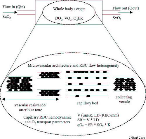
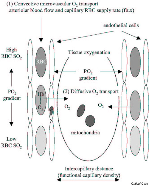
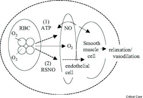
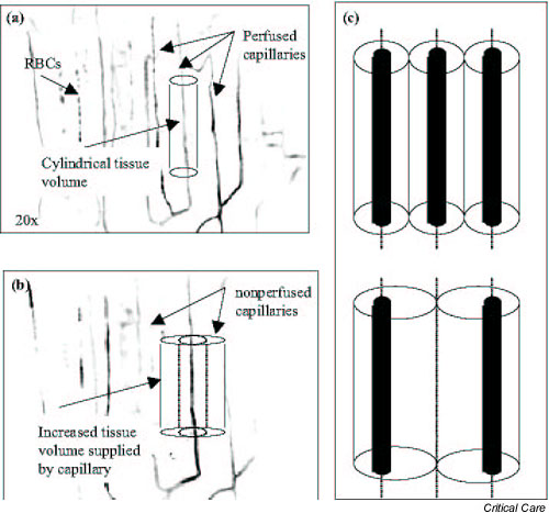
The microcirculation is a complex and integrated system that supplies and distributes oxygen throughout the tissues. The red blood cell (RBC) facilitates convective oxygen transport via co-operative binding with hemoglobin. In the microcirculation oxygen diffuses from the RBC into neighboring tissues, where it is consumed by mitochondria. Evidence suggests that the RBC acts as deliverer of oxygen and 'sensor' of local oxygen gradients. Within vascular beds RBCs are distributed actively by arteriolar tone and passively by rheologic factors, including vessel geometry and RBC deformability. Microvascular oxygen transport is determined by microvascular geometry, hemodynamics, and RBC hemoglobin oxygen saturation. Sepsis causes abnormal microvascular oxygen transport as significant numbers of capillaries stop flowing and the microcirculation fails to compensate for decreased functional capillary density. The resulting maldistribution of RBC flow results in a mismatch of oxygen delivery with oxygen demand that affects both critical oxygen delivery and oxygen extraction ratio. Nitric oxide (NO) maintains microvascular homeostasis by regulating arteriolar tone, RBC deformability, leukocyte and platelet adhesion to endothelial cells, and blood volume. NO also regulates mitochondrial respiration. During sepsis, NO over-production mediates systemic hypotension and microvascular reactivity, and is seemingly protective of microvascular blood flow.


Figures

Figure 1
Figure 1
Schematic representation of global and microvascular oxygen (O2) transport parameters. Arteriolar tone establishes the blood flow into an organ, and capillary resistance and rheology factors determine red blood cell (RBC) distribution (heterogeneity) within the capillary bed. Capillary hemodynamics are quantified as RBC velocity (V), lineal density (LD), and supply rate (SR) or flux. Capillary O2 transport (qO2) is determined by capillary SR, RBC hemoglobin O2 saturation (SO2), and the O2 carrying capacity of the RBC (K).
Figure 2
Figure 2
Schematic representation of convective and diffusive oxygen (O2) transport in the microcirculation. O2 is carried by the red blood cell (RBC; convective transport) from the lung microcirculation to the tissue microcirculation. As the RBC traverses the vascular bed it 'offloads' O2 to the neighboring tissue; O2 then diffuses from the capillary to the tissue mitochondria, where it is consumed. Local oxygen tension (PO2) gradients are established along the capillary vessel, as the RBC hemoglobin (Hb) O2 saturation (SO2) decreases, and into the tissue with the latter acting as the driving force of O2 diffusion.
Figure 3
Figure 3
The red blood cell (RBC) as a deliverer and 'sensor' of oxygen (O2). By altering its conformation, hemoglobin facilitates the release of nitrosothiol (RSNO; nitric oxide [NO] derivative) or ATP in response to increased O2 tension (PO2) gradients or mechanical deformation. Both RSNO and ATP evoke vasodilation, with the latter being NO mediated.
Figure 4
Figure 4
Sepsis induced capillary stopped-flow and increased effective tissue volume. (a, b) Consecutive microvascular variance images of the same rat skeletal muscle capillary bed at different times during the progression of sepsis. (Variance images depict the change in light intensity at each pixel in the field of view, from a 30 s video sequence. Perfused capillaries appear as dark lines, and the tissue background is white.) Sepsis increased capillary stopped-flow, reduced functional capillary density, and increased the effective tissue volume supplied by the remaining vessels. The latter is depicted in (c). RBC, red blood cell.
Figure 5
Figure 5
Relationships between global hemodynamics, tissue oxygen tension (PO2), and skeletal muscle lactate and ATP. In a 6-hour rat model of sepsis (cecal ligation and perforation [CLP]), fluid resuscitation was found to prevent decreases in systemic oxygen delivery (DO2) and tissue PO2 in skeletal muscle (tPO2). This apparent rescue of the muscle microcirculation was belied by elevated lactate and reduced ATP. *P < 0.05, versus control. FR, albumin fluid resuscitation; MAP, mean arterial pressure. By permission from Circ Shock 1988, 26:311–20 [35].
Figure 6
Figure 6
Local capillary oxygen extraction ratio (O2ERc) versus regional capillary stopped-flow (%CDstop). In a 24-hour rat model of sepsis (cecal ligation and perforation [CLP]), capillary oxygen extraction from normally flowing capillaries (capillary velocity between 20 and 325 μm/s) was found to correlate with the degree of regional capillary stopped-flow. As capillary stopped-flow increased, the RBCs offloaded increased amounts of oxygen. (CLP group: y = 0.018x - 0.18; r2= 0.64; P < 0.05.) By permission from Am J Physiol Heart Circ Physiol 2002, 282:H156–H164 [30].
Figure 7
Figure 7
Dependence on oxygen concentration of the percentage of inhibition of cytochrome c oxidase by nitric oxide (NO). For equal concentrations of cytochrome c oxidase and NO (1.2–1.8 μm) oxygen was found to compete with NO when the enzyme enters turnover. Each data point represents a different experimental setup. The results suggest that, under physiologic conditions, in which oxygen concentration is low, nanomolar concentrations of NO can regulate mitochondrial respiration. By permission from Biochem J 1995, 312:169–173 [140]. © The Biochemical Society.

References

    1. Gilbert RP. Mechanisms of the hemodynamic effects of endotoxin. Physiol Rev. 1960;40:245–278. - PubMed
    1. Carroll GC, Snyder JV. Hyperdynamic severe intravascular sepsis depends on fluid administration in cynomolgus monkey. Am J Physiol. 1982;243:R131–R141. - PubMed
    1. Krishnagopalan S, Kumar A, Parrillo JE, Kumar A. Myocardial dysfunction in the patient with sepsis. Curr Opin Crit Care. 2002;8:376–388. - PubMed
    1. Zhang H, Rogiers P, Smail N, Cabral A, Preiser JC, Peny MO, Vincent JL. Effects of nitric oxide on blood flow distribution and O2 extraction capabilities during endotoxic shock. J Appl Physiol. 1997;83:1164–1173. - PubMed
    1. Yang S, Cioffi WG, Bland KI, Chaudry IH, Wang P. Differential alterations in systemic and regional oxygen delivery and consumption during the early and late stages of sepsis. J Trauma. 1999;47:706–712. - PubMed

Publication types