. 2003 Sep 15;17(18):2334-45.
doi: 10.1101/gad.1121403.

Phage N4 RNA polymerase II recruitment to DNA by a single-stranded DNA-binding protein

Affiliations

Phage N4 RNA polymerase II recruitment to DNA by a single-stranded DNA-binding protein

Richard H Carter et al. Genes Dev. .

Abstract

Transcription of bacteriophage N4 middle genes is carried out by a phage-coded, heterodimeric RNA polymerase (N4 RNAPII), which belongs to the family of T7-like RNA polymerases. In contrast to phage T7-RNAP, N4 RNAPII displays no activity on double-stranded templates and low activity on single-stranded templates. In vivo, at least one additional N4-coded protein (p17) is required for N4 middle transcription. We show that N4 ORF2 encodes p17 (gp2). Characterization of purified gp2revealed that it is a single-stranded DNA-binding protein that activates N4 RNAPII transcription on single-stranded DNA templates through specific interaction with N4 RNAPII. On the basis of the properties of the proteins involved in N4 RNAPII transcription and of middle promoters, we propose a model for N4 RNAPII promoter recognition, in which gp2plays two roles, stabilization of a single-stranded region at the promoter and recruitment of N4 RNAPII through gp2-N4 RNAPII interactions. Furthermore, we discuss our results in the context of transcription initiation by mitochondrial RNA polymerases.


Figures

Figure 2.
Figure 2.
(A) p17 is encoded by ORF2. 35S-labeled proteins were separated by electrophoresis on SDS-PAGE and detected by autoradiography. (Lanes 1-3) Whole cell lysate from W3350(DE3)/pLysS/pSH2. (Lane 1) Uninduced cells. (Lane 2) IPTG-induced cells. (Lane 3) IPTG-induced and rifampicin-pretreated cells. (Lane 4) Rifampicinpretreated cells infected with wild-type phage. (Lane 5) Rifampicin-pretreated cells infected with N4am98 phage. Arrow indicates the position of p17. (B) Recombinant p17 is functional in vivo. Time course of DNA synthesis: (○) W3350cells infected with N4 wild-type phage; (•) W3350cells infected with N4 am98 phage; (□) W3350 (DE3)/pLysS/pSH2 cells pretreated with IPTG 15 min prior to infection with N4am98 phage. (C) Gp2 purification. Protein fractions from different steps of the purification are shown. (Lane 1) A 25% ammonium sulfate supernatant. (Lane 2) Solubilized 50% ammonium sulfate pellet. (Lane 3) Sephacryl S-300 HR eluate. (Lane 4) Hydroxylapatite eluate. (Lane 5) Q-Sepharose eluate. (Lane 6) Final dialysate.
Figure 1.
Figure 1.
Sequence of the p17-coding region reveals a 128-amino acid ORF. The N4am98 mutation, a CAG (Gln, amino acid 58)-to-TAG (amber) transition, is bold. The tryptophan (amino acid 30) is boxed.
Figure 3.
Figure 3.
Gp2 binds to single-stranded DNA. (A) EMSA of labeled template and complementary strands of the 171-nucleotide Mc fragment (1 nM) and increasing concentrations of gp2. Concentrations of gp2 are as follows: none (lanes 1,10); 8 nM (lanes 2,11); 16 nM (lanes 3,12); 31 nM (lanes 4,13); 62 nM (lanes 5,14); 125 nM (lanes 6,15); 0.25 μM (lanes 7,16); 0.5 μM (lanes 8,17); 1 μM (lanes 9,18). (B) EMSA of reactions containing a labeled 171 nucleotide Mc complementary strand (1 nM) incubated with the indicated amount of nucleic acid competitor prior to addition of gp2. (dsDNA) pBR[Mc]; (RNA) CsCl-purified RNA from N4-infected cells; (ssDNA) M13 mp18 viral DNA. (Lane 1) Free probe. (Lane 2) Probe and gp2. Competitor amounts are as follows: 0.05 ng (lanes 3,9,15); 0.5 ng (lanes 4,10,16); 5 ng (lanes 5,11,17); 50ng (lanes 6,12,18); 0.5 μg (lanes 7,13,19); 5 μg (lanes 8,14,20).
Figure 4.
Figure 4.
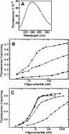
Analysis of gp2-nucleic acid binding by fluorescence quenching. (A) Fluorescence spectrum of gp2. Gp2 was present at 250mM in fluorescence buffer containing 300nM NaCl. (B) Gp2 binds preferentially to pyrimidine-containing oligonucleotides. A total of 30base homopolymers were titrated into a solution containing 0.5 μM gp2. (•) [dT]30; (▴) [dC]30; (▪) [dA]30. (C) Dependence of gp2 binding on oligonucleotide length. Poly-[dT] oligonucleotides were titrated into a solution containing 0.25 μM gp2 and 300 mM NaCl. (•) 20 mer; (▴) 30 mer; (▪) 40 mer; (▾) 50mer.
Figure 5.
Figure 5.
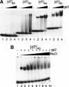
(A) Effect of oligonucleotide length on gp2 binding to ssDNA. Reactions contained 50nM oligonucleotide and gp2 in 300mM NaCl, 1 mM EDTA, 20mM Tris-Cl (pH 8.0). Concentrations of gp2 are as follows: none (lane 1); 0.25 μM (lane 2); 0.50 μM (lane 3); 1 μM (lane 4). (B) Effect of NaCl concentration on gp2 binding to ssDNA. Reactions contained 50nM [dT]40 and 0.75 μM gp2. Gp2 was preincubated for 5 min at the indicated NaCl concentration in 1 mM EDTA, 20mM Tris-Cl (pH 8.0), and the oligonucleotide was added 5 min prior to loading of the gel. Lanes 1 and 2 contained no NaCl; lanes 3-10 contained 0.25, 0.5, 0.75, 1.0, 1.25, 1.5, 1.75, and 2.0 M NaCl, respectively.
Figure 6.
Figure 6.
(A) Gp2 activates N4 RNAPII transcription on single-stranded templates. In vitro transcription reactions contained 50nM of the 171-nucleotide template or complementary strands of the N4 Mc fragment, 40nM RNAPII, and increasing amounts of gp2. The positions of DNA molecular weight markers are indicated on the left. Asterisk denotes transcript initiating close to in vivo start site and arrows denote major transcripts. (B) Transcription initiates at the same sites in the absence or presence of gp2. S1 nuclease protection analysis of RNA products synthesized from the N4 Mc complementary strand. Template DNA was incubated in the absence or presence of 1.0μM gp2 and 40nM RNAPII for 10min. RNA products were isolated and hybridized to the end-labeled oligonucleotide MCC101-165. Only 12% of the RNA synthesized in the presence of gp2 was used, to allow comparison of the start sites. (Right lanes) Maxam-Gilbert sequencing reactions of the end-labeled probe. The positions of DNA molecular weight markers are indicated on the left. (C) Gp2 does not activate transcription at high RNAPII concentrations. Transcription reactions were carried out with the indicated amount of RNAPII and 50 nM MCC101-165 DNA in the absence or presence of 0.4 μM gp2. The positions of DNA molecular weight markers are indicated on the left.
Figure 7.
Figure 7.
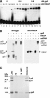
Analysis of gp2-N4 RNAPII complex formation by gel retardation and Western blotting. (A) Increasing gp2 or RNAPII concentrations stimulates complex formation on single-stranded template. Reactions containing 50nM end-labeled MCC101-165 ssDNA, gp2, and RNAPII in transcription buffer were incubated for 15 min at 4°C. (B) Gp2 and RNAPII are present in a complex on ssDNA. Gp2, recombinant hexahistidine-tagged RNAPII, or a mixture of both proteins was incubated in the absence or presence of MCC101-165 ssDNA. Complexes were separated on native gels and probed with anti-gp2 antibody (left) or Xpress antibody (right). Asterix denotes RNAPII-gp2 complex formed in the absence of ssDNA. (C) Gp2 interacts with RNAPII. Recombinant hexahistidine-tagged RNAPII was incubated with gp2 and applied to a metal-affinity column.

Similar articles

Cited by

References

    1. Abravaya K. and Rothman-Denes, L.B. 1989a. In vitro requirements for N4 RNA polymerase II-specific initiation. J. Biol. Chem. 264: 12695-12699. - PubMed
    1. ____. 1989b. N4 RNA polymerase II sites of transcription initiation. J. Mol. Biol. 211: 359-372. - PubMed
    1. Ausubel F.M., Brent, R., Kingston, R.E., Moore, D.D., Seidman, J.G., Smith, J.A., and Struhl, K. (eds.) 1999. Current protocols in molecular biology. Wiley, New York, N.Y.
    1. Blackwell L. and Borowiec, J. 1994. Human replication protein A binds single-stranded DNA in two distinct complexes. Mol. Cell. Biol. 14: 3993-4001. - PMC - PubMed
    1. Bochkareva E., Belegu, V., Korolev, S., and Bochkarev, A. 2001. Structure of the major single-stranded DNA-binding domain of replication protein A suggests a dynamic mechanism for DNA binding. EMBO J. 20: 612-618. - PMC - PubMed

Publication types

MeSH terms