. 2003 Oct 1;17(19):2356-61.
doi: 10.1101/gad.1131103. Epub 2003 Sep 15.

The budding yeast silencing protein Sir1 is a functional component of centromeric chromatin

Affiliations

The budding yeast silencing protein Sir1 is a functional component of centromeric chromatin

Judith A Sharp et al. Genes Dev. .

Abstract

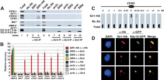
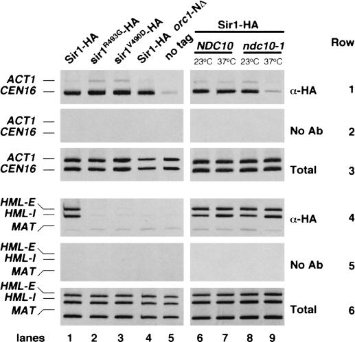
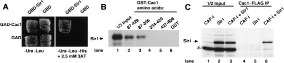
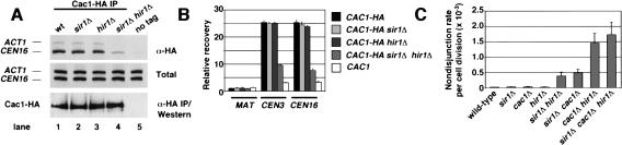
In fission yeast and multicellular organisms, centromere-proximal regions of chromosomes are heterochromatic, containing proteins that silence gene expression. In contrast, the relationship between heterochromatin proteins and kinetochore function in the budding yeast Saccharomyces cerevisiae remains largely unexplored. Here we report that the yeast heterochromatin protein Sir1 is a component of centromeric chromatin and contributes to mitotic chromosome stability. Sir1 recruitment to centromeres occurred through a novel mechanism independent of its interaction with the origin recognition complex (ORC). Sir1 function at centromeres was distinct from its role in forming heterochromatin, because the Sir2-4 proteins were not associated with centromeric regions. Sir1 bound to Cac1, a subunit of chromatin assembly factor I (CAF-I), and helped to retain Cac1 at centromeric loci. These studies reveal that although budding yeast and mammalian cells use fundamentally different mechanisms of forming heterochromatin, they both use silencing proteins to attract the histone deposition factor CAF-I to centromeric chromatin.


Figures

Figure 1.
Figure 1.
Association of Sir1 with centromeric chromatin. (A) Presence of Sir1, but not Sir2-4, at centromeric loci. Formaldehyde cross-linked chromatin prepared from yeast strains CFY416 (SIR1-HA; lanes 2,4) and PKY346 (SIR1; lane 3) was immunoprecipitated in the presence of monoclonal 12CA5 anti-HA antibody (lanes 3-4) or mock treated (lane 2). Chromatin from yeast strains PKY090 (wild-type SIR2, SIR3, SIR4), PKY3342 (sir2Δ), PKY3343 (sir3Δ), and PKY3344 (sir4Δ) was mock treated (lane 6), or immunoprecipitated with antibodies to Sir2 (lanes 7,8), Sir3 (lanes 9,10), and Sir4(lanes 11,12). PCR was performed to visualize recovery of the core centromeric regions of CEN3, CEN11, CEN16, the HMR-E silencer, MAT, ACT1, and two subtelomeric sequences on the right arm of Chromosome VI (TEL). Total chromatin was titrated to determine the linear range of the PCR (data not shown); a 1:32 dilution that falls within this range is shown in lanes 1 and 5. (B) Quantitation of Sir1-4 chromatin immunoprecipitation experiments. Chromatin immunoprecipitations (n = 3 for each genotype) were performed as described in A. The signal strength of PCR products was measured using Quantity One software (Bio-Rad) and used to calculate the percent recovery of HMR-E, MAT, CEN1-4, CEN11, and CEN16 PCR products. The average percent recovery is expressed as fold enrichment relative to the MAT control locus. (C) Distribution of Sir1 across the CEN3 region. Chromatin was prepared from yeast strain CFY416 (SIR1-HA), and PCR was performed as in A. Anti-HA-precipitated chromatin, mock-precipitated chromatin, and total chromatin was analyzed for recovery of fragments at or flanking the core centromeric region of CEN3 as indicated on the diagram (not to scale). (D) Sir1 colocalizes with centromere protein Ndc10. Yeast strain PKY2648 (SIR1-HA, NDC10-GFP) was prepared for indirect immunofluorescence analysis as described (Loidl et al. 1998). Spread nuclei were stained with anti-HA (red), anti-GFP (green) antibodies, and DAPI (blue). Colocalization of Sir1 and Ndc10 is indicated by the yellow staining in the merged image.
Figure 2.
Figure 2.
Sir1 association with centromeric chromatin requires Ndc10, but is independent of the Orc1 N terminus. Chromatin immunoprecipitation was performed as described in Figure 1. Total (rows 3,6), mock immunoprecipitations (rows 2,5), and anti-HA immunoprecipitations (rows 1,4) were then tested for the presence of CEN16, ACT1, HML-E, HML-I, and MAT DNA. Yeast strains were PKY2586 (SIR1-HA; lanes 1,6,7), CFY687 (sir1R493G-HA; lane 2), CFY689 (sir1V490D-HA; lane 3), CFY1392 (SIR1-HA, orc1-NΔ; lane 4), CFY345 (SIR1; lane 5), and PKY2578 (SIR1-HA, ndc10-1; lanes 8,9).
Figure 3.
Figure 3.
Biochemical interaction between Sir1 and Cac1. (A) Two-hybrid interaction in yeast. Yeast strain PJ69-4a (James et al. 1996) expressed the indicated activation domain (GAD) or DNA-binding (GBD) fusion proteins. The GAD-Cac1 plasmid encodes amino acids 217-606 of Cac1, and was recovered in a two-hybrid screen using the full-length GBD-Sir1 plasmid. Cells were grown either on media lacking uracil and leucine (-Ura -Leu) to select for the two plasmids or on -Ura -Leu media also lacking histidine and containing 2.5 mM 3-aminotriazole (-His + 3AT) to score for activation of the HIS3 reporter gene, indicating interaction between the fusion proteins. (B) For direct interaction in vitro, 2.5 μg of unfused GST (lane 6) or GST-Cac1 fusion proteins [Krawitz et al. 2002; Cac1 amino acids 87-429 (lane 2), 87-306 (lane 3), 334-429 (lane 4), or 429-606 (lane 5)] prebound to 15 μL of Glutathione Agarose (Sigma) were incubated with 6 μL of 35S-labeled in vitro translated Sir1 (Novagen). Reactions were rotated at 4°C for 1 h prior to three 1-mL washes with Buffer A (25 mM Tris-HCl at pH 7.5, 1 mM EDTA, 0.1% NP-40) plus 250 mM NaCl. Bound proteins were eluted with SDS sample buffer, separated by SDS-PAGE, and detected by autoradiography. Lane 1 contains 2 μL of in vitro translated Sir1 loaded directly onto the gel. (C) Interaction between CAF-I and Sir1 in cell extracts. Overproduction of CAF-I including a Flag-tagged Cac1 subunit and Sir1 in SF9 cells was performed as described (Sharp et al. 2001). Five microliters of nuclear extract containing CAF-I (lane 4), Sir1 (lane 5), or both CAF-I and Sir1 (lane 6) were incubated with 95 μL of Buffer A plus 50 mM NaCl at 4°C for 2 h in the presence of anti-Flag antibodies (Sigma) cross-linked to Protein A-Sepharose. Samples were washed three times with 1 mL of Buffer A plus 500 mM NaCl. Bound proteins were eluted with SDS sample buffer, separated by SDS-PAGE, and analyzed by immunoblotting with a polyclonal anti-Sir1 antibody. Also, 2.5 μL of total nuclear extract from cells expressing CAF-I alone (lane 1), Sir1 alone (lane 2), or both CAF-I and Sir1 (lane 3) were analyzed on the same gel. The asterisk indicates a cross-reacting protein present in the crude cell lysates.
Figure 4.
Figure 4.
Cac1, Hir1, and Sir1 make overlapping contributions to centromere structure and function. (A) Cac1 association with the core centromeric region requires Sir1 and Hir1. Chromatin immunoprecipitations were performed on yeast strains YB703 (CAC1-HA; lane 1), PKY2615 (CAC1-HA, sir1Δ; lane 2), PKY2617 (CAC1-HA, hir1Δ; lane 3), PKY2619 (CAC1-HA, sir1Δ, hir1Δ; lane 4), and PKY346 (CAC1; lane 5). Total, mock (data not shown), and anti-HA immunoprecipitations were then tested for the presence of CEN16 and ACT1 DNA. A Western blot was performed to compare immunoprecipitation of Cac1-HA from the indicated strains (bottom panel). (B) Quantitation of Cac1-HA chromatin immunoprecipitation experiments. Chromatin immunoprecipitations (n = 3 for each genotype) were analyzed as described in Figure 1B for the MAT, CEN3, and CEN16 loci. (C) SIR1, HIR1, and CAC1 together prevent chromosomal nondisjunction during mitotic cell divisions. Nondisjunction rates per cell division were quantified in haploid yeast strains containing a nonessential chromosome fragment by the half-sector colony color assay as described (Shero et al. 1991; Sharp et al. 2002). Yeast strains are PKY847 (wild-type), PKY2609 (sir1Δ), PKY2216 (cac1Δ), PKY865 (hir1Δ), PKY2655 (sir1Δ hir1Δ), PKY 2610 (sir1Δ cac1Δ), PKY 2217 (cac1Δ hir1Δ), and PKY2656 (sir1Δ cac1Δ hir1Δ). Three experiments for each genotype (except PKY2656, n = 4) were performed; the average chromosome nondisjunction rate per cell division and standard deviation (error bars) are shown.

Similar articles

Cited by

References

    1. Bernard P., Maura, J.F., Partridge, J.F., Genier, S., Javerzat, J.P., and Allshire, R.C. 2001. Requirement of heterochromatin for cohesion at centromeres. Science 294: 2539-2542. - PubMed
    1. Biggins S. and Walczak, C.E. 2003. Captivating capture: How microtubules attach to kinetochores. Curr. Biol. 13: R449-R460. - PubMed
    1. Briggs S.D., Bryk, M., Strahl, B.D., Cheung, W.L., Davie, J.K., Dent, S.Y., Winston, F., and Allis, C.D. 2001. Histone H3 lysine 4 methylation is mediated by Set1 and required for cell growth and rDNA silencing in Saccharomyces cerevisiae. Genes & Dev. 15: 3286-3295. - PMC - PubMed
    1. Bryk M., Briggs, S.D., Strahl, B.D., Curcio, M.J., Allis, C.D., and Winston, F. 2002. Evidence that Set1, a factor required for methylation of histone H3, regulates rDNA silencing in S. cerevisiae by a Sir2-independent mechanism. Curr. Biol. 12: 165-170. - PubMed
    1. Chen Y., Baker, R.E., Keith, K.C., Harris, K., Stoler, S., and Fitzgerald-Hayes, M. 2000. The N terminus of the centromere H3-like protein Cse4p performs an essential function distinct from that of the histone fold domain. Mol. Cell. Biol. 20: 7037-7048. - PMC - PubMed

Publication types

MeSH terms

Substances