. 2003 Sep 15;162(6):963-9.
doi: 10.1083/jcb.200305082.

Centrosome positioning in interphase cells

Affiliations

Centrosome positioning in interphase cells

Anton Burakov et al. J Cell Biol. .

Abstract

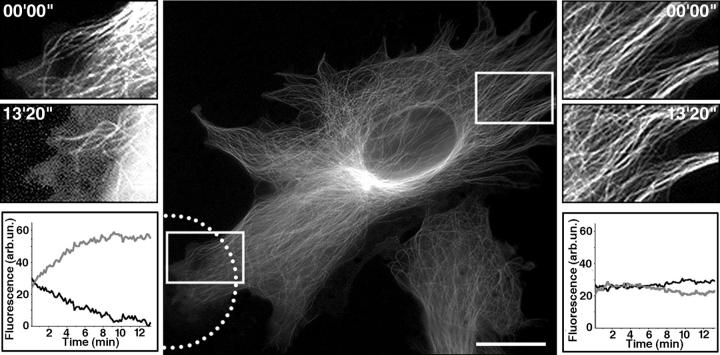
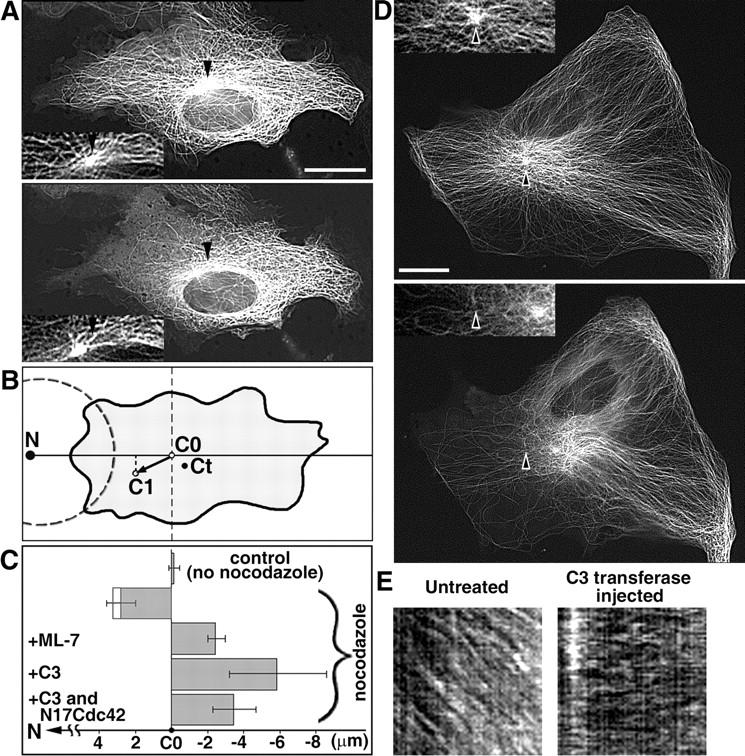
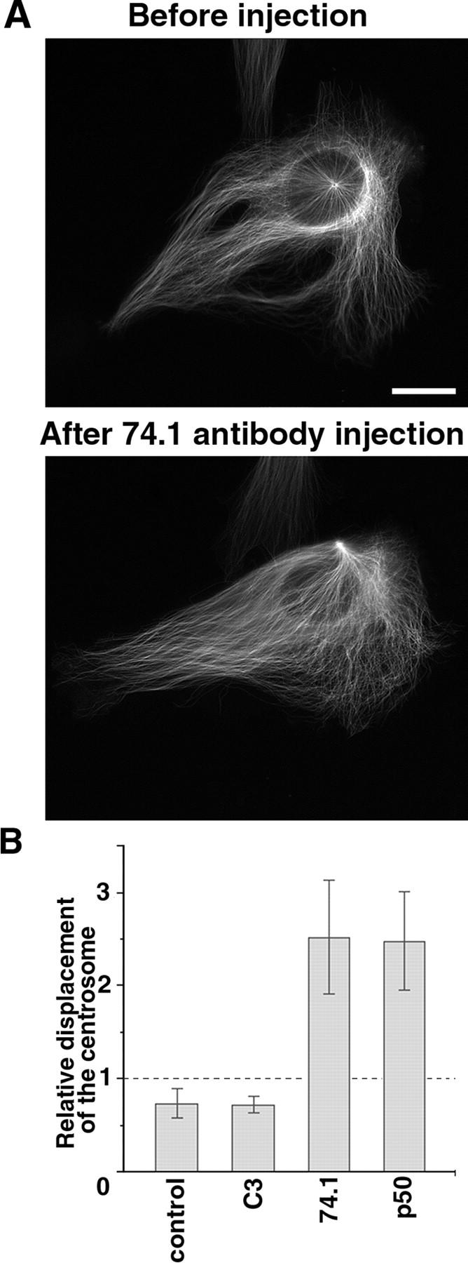
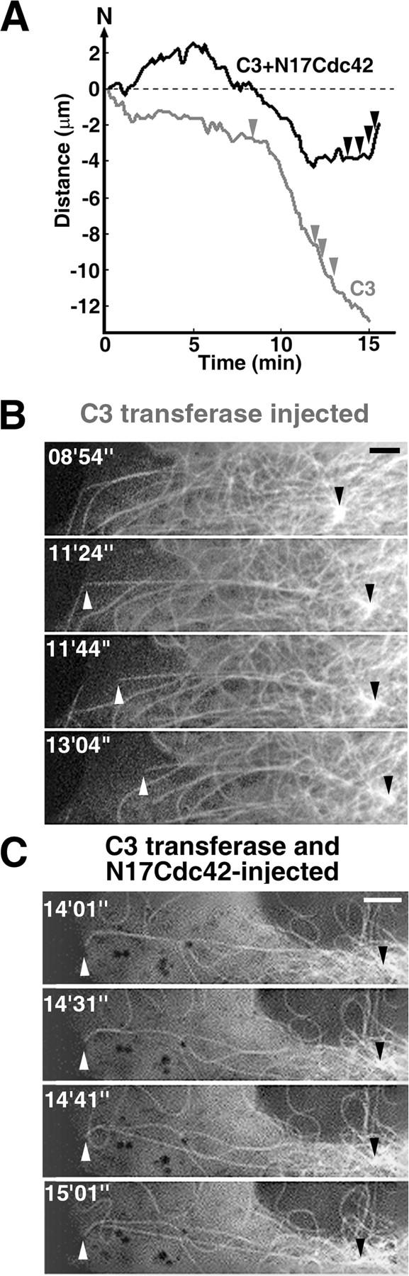
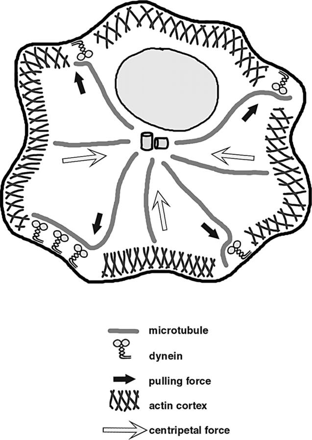
The position of the centrosome is actively maintained at the cell center, but the mechanisms of the centering force remain largely unknown. It is known that centrosome positioning requires a radial array of cytoplasmic microtubules (MTs) that can exert pushing or pulling forces involving MT dynamics and the activity of cortical MT motors. It has also been suggested that actomyosin can play a direct or indirect role in this process. To examine the centering mechanisms, we introduced an imbalance of forces acting on the centrosome by local application of an inhibitor of MT assembly (nocodazole), and studied the resulting centrosome displacement. Using this approach in combination with microinjection of function-blocking probes, we found that a MT-dependent dynein pulling force plays a key role in the positioning of the centrosome at the cell center, and that other forces applied to the centrosomal MTs, including actomyosin contractility, can contribute to this process.


Figures

Figure 1.
Figure 1.
Local disruption of MTs in a cell by the local application of nocodazole. (Center) low magnification live fluorescence image of a cell with MTs labeled by injecting fluorescently tagged tubulin subunits. Image was obtained just before application through the micropipette of a nocodazole solution in the area depicted by the dashed line. Micrographs on the left and right are high magnification images of MTs in the boxed regions shown in the central panel acquired at the cell edge proximal to (left) or distal from (right) the micropipette tip, before (top) or after (bottom) the application of nocodazole. Time-lapsed series of MT dynamics corresponding to the micrographs on left and right are shown in Videos 1 and 2, available at http://www.jcb.org/cgi/content/full/jcb.200305082/DC1. The graphs show kinetics of changes in the levels of tubulin monomer (plots shown in gray) or polymer (plots shown in black) at the cell edges proximal (left) or distal (right) to the micropipette tip. Bar, 20 μm.
Figure 2.
Figure 2.
Centrosome position is maintained through a MT mediated pulling force. (A and D) Pairs of live fluorescence images of the centrosome acquired before (top image of each pair) and after (bottom image of each pair) the local application of nocodazole. Insets show the position of the centrosome in the same cells at higher magnification. The initial positions of the centrosome are indicated by black arrows. See also the corresponding movies (Videos 3 and 4) for full time-lapse series, available at http://www.jcb.org/cgi/content/full/jcb.200305082/DC1. (A) Noninjected cell. Local application of nocodazole induces centrosome movement toward the application site. (D) Cell injected with a Rho inhibitor C3 transferase (0.1 mg/ml). Inhibition of RhoA activity reverses the direction of the centrosome movement upon application of nocodazole. (B) Method for the quantification of the centrosome movement. The centrosome position (C) was determined as the focus of the MT fluorescence. Centroid position (Ct) was calculated by Metamorph software as the point equidistant from all the cell margins. Centrosome movement was calculated as the distance between the initial (C0) and final (C1) positions of the centrosome, projected onto a straight line connecting the nocodazole pipette tip (N) and the centrosome at time point zero. Observations were made in stationary cells, but cell shape changes during the time of the experiment (6–15 min) sometimes led to small changes in the calculated position of the centroid. In such cases, centroid displacement was projected onto the same straight line and the vector sum of the centroid, and centrosome displacement was calculated to obtain the final value of the centrosome movement. (C) Quantification of the centrosome movement. Positive value of movement was considered when the centrosome moved from the initial position (C0) toward the nocodazole pipette tip (N). Open bar shows centrosome displacement in cytoplasts. (E) Kymograph analysis of rhodamine F-actin speckles in control (left) and C3-transferase–injected (right) cell. The absence of the centripetal flow after the injection of C3-transferase is evident from the lack of directional movement of the fluorescent speckles (right), compared with the diagonal pattern of movement in the control cells (left). Bars, 20 μm.
Figure 3.
Figure 3.
Positioning of the centrosome in the cell center requires the activity of cytoplasmic dynein. (A) Live fluorescence images of MTs in a cell before (top) or 45 min after (bottom) the injection of a dynein blocking antibody 74.1. After the injection of dynein blocking antibody, the centrosome moved to the cell margin. (B) Quantification of the displacement of the centrosome in cells injected with C3 transferase, antibody 74.1, or the p50 subunit of dynactin. Relative displacement of the centrosome was determined as the ratio between the initial (before injection) and the final (after injection) positions of the centrosome, calculated as the percentage of the cell radius drawn from the centroid through the centrosome. In control and C3-injected cells, where the centrosome remained in place, the ratios were close to 1. In cells injected with antibody 74.1 or the p50 subunit of dynactin, the centrosome traveled significantly toward the nearest cell margin, causing the greatly increased displacement values. Bar, 20 μm.
Figure 4.
Figure 4.
Centrosome positioning is regulated by Cdc42-dependent signaling pathway. (A) Kinetics of the centrosome movement in cells injected with Rho inhibitor C3 transferase (gray), or a combination of C3-transferase and Cdc42 dominant–negative recombinant protein N17Cdc42 (black). Arrowheads on the curves show the time points where images were taken in B and C. (B and C) Time series of the fluorescence images of MTs showing centrosome movement quantified in A, injected with C3 transferase (B) or double injected with C3 transferase and N17Cdc42 (C). See also the corresponding movies (Videos 5 and 6) for full time-lapse series, available at http://www.jcb.org/cgi/content/full/jcb.200305082/DC1. (B) In the absence of actin centripetal flow, the centrosome (black arrowhead) moves continuously away from the nocodazole pipette tip, driven by the pulling force on the MTs at the cortex. The pulling force on the end distal to the pipette tip is strong enough to induce breakage of the MTs at the proximal end (white arrowhead shows the position of the nascent end of the broken MT), accelerating the centrosome movement away from the pipette tip (as shown in A). (C) In the absence of both actin centripetal flow and the activity of Cdc42, centrosome moves back and forth around the central point. Pulling forces applied at the cortex are not enough to induce MT breakage. A single MT is enough to anchor the centrosome and pull it back toward the pipette tip, causing reversal of the direction of the movement (as shown in A). White arrowhead shows the position of the anchored MT end. Bars, 5 μm.
Figure 5.
Figure 5.
Forces involved in the positioning of the centrosome. Pulling forces applied to the MTs at the cell cortex by dynein act to position the centrosome at the cell center. Pushing forces, including actin centripetal flow and MT dynamics, are directed toward the cell center.

Similar articles

Cited by

References

    1. Burkhardt, J.K., C.J. Echeverri, T. Nilsson, and R.B. Vallee. 1997. Overexpression of the dynactin complex disrupts dynein-dependent maintenance of membrane organelle distribution. J. Cell Biol. 139:469–484. - PMC - PubMed
    1. Cramer, L.P. 1997. Molecular mechanism of actin-dependent retrograde flow in lamellipodia of motile cells. Front. Biosci. 2:d260–d270. - PubMed
    1. Dillmann, J.F., III, and K. Pfister. 1994. Differential phosphorylation in vivo of cytoplasmic dynein associated with anterogradely moving organelles. J. Cell Biol. 127:1671–1681. - PMC - PubMed
    1. Dogterom, M., and B. Yurke. 1997. Measurement of force-velocity relation for growing microtubules. Science. 278:856–860. - PubMed
    1. Dujardin, D.L., and R.B. Vallee. 2002. Dynein at the cortex. Curr. Opin. Cell Biol. 14:44–49. - PubMed

Publication types

MeSH terms