. 2003 Sep 15;162(6):1123-33.
doi: 10.1083/jcb.200302090.

Site-specific inductive and inhibitory activities of MMP-2 and MMP-3 orchestrate mammary gland branching morphogenesis

Affiliations

Site-specific inductive and inhibitory activities of MMP-2 and MMP-3 orchestrate mammary gland branching morphogenesis

Bryony S Wiseman et al. J Cell Biol. .

Abstract

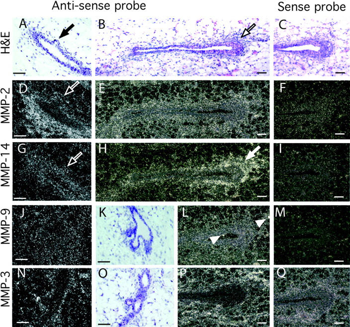
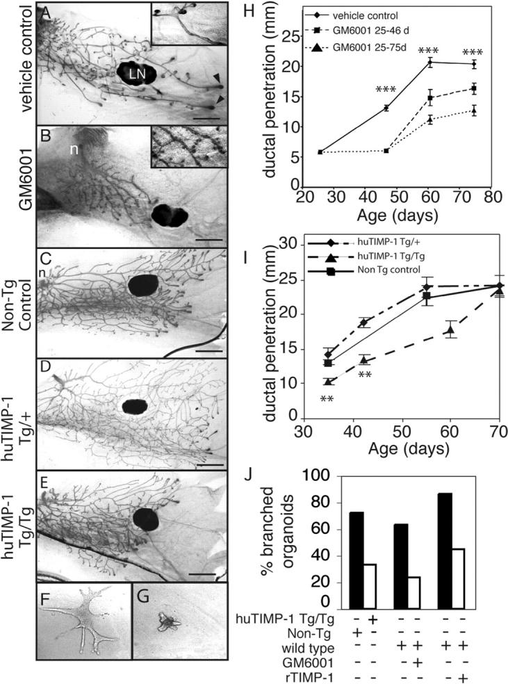
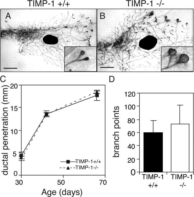
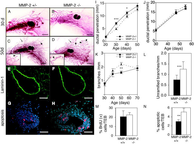
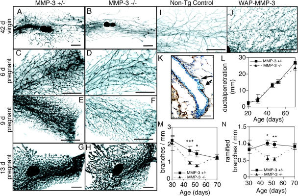
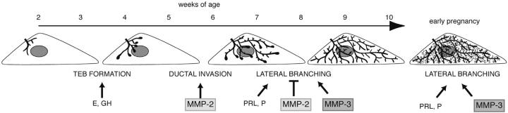
During puberty, mouse mammary epithelial ducts invade the stromal mammary fat pad in a wave of branching morphogenesis to form a complex ductal tree. Using pharmacologic and genetic approaches, we find that mammary gland branching morphogenesis requires transient matrix metalloproteinase (MMP) activity for invasion and branch point selection. MMP-2, but not MMP-9, facilitates terminal end bud invasion by inhibiting epithelial cell apoptosis at the start of puberty. Unexpectedly, MMP-2 also represses precocious lateral branching during mid-puberty. In contrast, MMP-3 induces secondary and tertiary lateral branching of ducts during mid-puberty and early pregnancy. Nevertheless, the mammary gland is able to develop lactational competence in MMP mutant mice. Thus, specific MMPs refine the mammary branching pattern by distinct mechanisms during mammary gland branching morphogenesis.


Figures

Figure 1.
Figure 1.
Localization of MMPs-2, -3, -9, and -14 mRNA within the mammary gland. Mammary glands were taken at 50 d old and sectioned. (A–C, K, and O) Hematoxylin and eosin counterstain of mammary gland sections in G–J and N, respectively. Note the initiating lateral branch in A (black arrow) and TEB in B (black outlined arrow). In situ hybridization analysis was performed with the following antisense and sense probes (as indicated): (D–F) MMP-2, (G–I) MMP-14, (J, L, and M) MMP-9, and (N, P, and Q) MMP-3. Note the reduction in MMP-2, but not MMP-14 mRNA, at the initiating lateral branch in the adjacent sections D and G (white outlined arrows); the localization of MMP-14 around the TEB in H (white arrow) and the spots of MMP-9 expression probably localized in macrophages in L (white arrow heads). Bars, 50 μm.
Figure 2.
Figure 2.
MMP inhibition disrupts mammary gland branching morphogenesis. (A and B) Mammary gland whole mounts from 6.5-wk-old mice treated daily with (A) vehicle or (B) GM6001 from 3.5 wk old. Note the extra ductal budding in the GM6001 treated mammary glands (inset). LN, lymph node; arrowheads, TEBs. Bar, 1 mm. (C–E) Mammary gland whole mounts from (C) nontransgenic control mice and (D) mice hemizygous and (E) homozygous for the β-actin human TIMP-1 transgene at 35 d old. n, nipple. Bar, 1 mm. (F and G) Primary mammary organoids grown for 7 d in the presence of EGF in collagen gels from (F) nontransgenic control mice or (G) huTIMP-1 transgenic mice. (H and I) Penetration of mammary ducts into fat pad of control mice, mice continually treated with GM6001 until sacrifice and mice that were treated with GM6001 from 3.5- to 6.5-wk-old using 8–12 mammary glands per data point (t test compared vehicle-control with mice continually treated with GM6001; H) and (I) nontransgenic control, Tg/+ and Tg/Tg hu-TIMP-1 transgenic mice using 4–12 mammary glands per data point. Data are mean ± SEM. t test compared Tg/+ with Tg/Tg. (J) The percentage of branched organoids in response to 7-d treatment with EGF. Organoids were derived from hu-TIMP-1 transgenic mice, nontransgenic controls, or wild-type mice and grown in absence or presence of GM6001 or recombinant TIMP-1 as indicated. In all panels: ***, P < 0.0005; **, P < 0.005, unpaired, two-tailed t test.
Figure 3.
Figure 3.
TIMP-1 is not necessary for ductal invasion or branching in the mammary gland. (A and B) Whole mounts of mammary glands of 42-d-old TIMP-1 +/+ (A) and TIMP-1 −/− mice (B). Note the enlarged TEBs of TIMP-1 −/− mice (inset). Bars, 1 mm. (C and D) Penetration of mammary ducts into fat pad (C) and number of branch points beyond the lymph node at 42 d old (D) of TIMP-1 +/+ and TIMP-1 −/− mice. Data are mean (C) ± SEM or (D) ± SD using four to eight mammary glands per data point.
Figure 4.
Figure 4.
MMP-2/mice have reduced ductal invasion and increased lateral branching. (A–D) Whole mounts of mammary glands of an (A and C) MMP-2 +/− and (B and D) MMP-2 −/− mice (A and B) 30 d old and (C and D) 50 d in estrus. Note supernumerary branching in D (inset). Example of ramified branch (arrows) and unramified branch or bud (arrowheads). Bars, 1 mm. (E–H) Sections through TEBs in mammary glands from 30-d-old (E and G) MMP-2 +/− and (F and H) MMP-2 −/− mice. (E and F) Immunohistochemistry for Ln-1. (G and H) TUNEL assay. Apoptotic cells are red. Bars, 25 μm. (I–L) Penetration of mammary ducts of (I) MMP-2 +/− and MMP-2 −/− and (J) MMP-9 +/− and MMP-9 −/− mice, (K) number of branches per millimeter, and (L) number of unramified branches per millimeter at 50 d from primary mammary ducts of MMP-2 +/− and −/− mice. Data are mean ± SEM (I, K, and L) using 8–16 or (H) 4–8 mammary glands per data point. (M and N) Percentage of BrdU (M) or TUNEL (N) positive cells within TEBs of MMP-2 −/− and MMP-2 +/− 30-d-old mice. Data are mean percent per TEB ± SD. ***, P < 0.0005; **, P < 0.005; *, P < 0.05, unpaired, two-tailed t test.
Figure 5.
Figure 5.
MMP-3 is required for lateral branching in the mammary gland. (A–H) Whole mounts of mammary glands of (A, C, E, and G) MMP-3 +/− and (B, D, F, and H) MMP-3 −/− mice at (A and B) 42 d old in estrus, (C and D) 6 d of pregnancy, (E and F) 9 d of pregnancy, and (G and H) 13 d of pregnancy. (I and J) Whole mounts of mammary glands of 70-d-old virgin (I) nontransgenic controls or (J) WAP-MMP-3 transgenic mice. Bars, 1 mm. (K) Wild-type mammary gland stained for Ln-1. Note reduction in Ln-1 where lateral branches are budding (arrows). Bar, 25 μm. (L–N) Penetration of ducts (L), number of total branches per millimeter (M), and number of ramified secondary branches per millimeter (N) from primary ducts of MMP-3 −/− and MMP-3 +/− mammary glands. Data are mean ± SEM using 4–12 mammary glands per data point. ***, P < 0.0005; **, P < 0.005; *P, < 0.05, unpaired, two-tailed t test.
Figure 6.
Figure 6.
Model for different phases of mammary gland branching morphogenesis. Before puberty, the mammary epithelial is small and simply branched. In response to the release of estrogen (E) and growth hormone (GH), at ∼3 wk old TEBs form. MMP-2 is then involved in inducing TEBs to invade and the ducts begin to fill the fat pad by branching dichotomously through bifurcation. At ∼6–8 wk old, the mammary ducts branch laterally. This process is suppressed by MMP-2 and induced by MMP-3 and may be related to changes in the response of the gland to progesterone (P) and prolactin (PRL). The fat pad is filled with ducts at ∼10 wk old and is relatively quiescent until pregnancy, when there is another wave of lateral branching that is regulated by MMP-3, P, and PRL before the formation of lobular alveoli.

Similar articles

Cited by

References

    1. Affolter, M., S. Bellusci, N. Itoh, B. Shilo, J.P. Thiery, and Z. Werb. 2003. Tube or not tube: remodeling epithelial tissues by branching morphogenesis. Dev. Cell. 4:11–18. - PubMed
    1. Alexander, C.M., E.W. Howard, M.J. Bissell, and Z. Werb. 1996. Rescue of mammary epithelial cell apoptosis and entactin degradation by a tissue inhibitor of metalloproteinases-1 transgene. J. Cell Biol. 135:1669–1677. - PMC - PubMed
    1. Alexander, C.M., S. Selvarajan, J. Mudgett, and Z. Werb. 2001. Stromelysin-1 regulates adipogenesis during mammary gland involution. J. Cell Biol. 152:693–703. - PMC - PubMed
    1. Baker, A.H., D.R. Edwards, and G. Murphy. 2002. Metalloproteinase inhibitors: biological actions and therapeutic opportunities. J. Cell Sci. 115:3719–3727. - PubMed
    1. Blatchford, D.R., L.H. Quarrie, E. Tonner, C. McCarthy, D.J. Flint, and C.J. Wilde. 1999. Influence of microenvironment on mammary epithelial cell survival in primary culture. J. Cell. Physiol. 181:304–311. - PubMed

Publication types

MeSH terms

Substances