. 2003 Sep 15;162(6):1161-72.
doi: 10.1083/jcb.200305078.

Association of TAG-1 with Caspr2 is essential for the molecular organization of juxtaparanodal regions of myelinated fibers

Affiliations

Association of TAG-1 with Caspr2 is essential for the molecular organization of juxtaparanodal regions of myelinated fibers

Maria Traka et al. J Cell Biol. .

Abstract

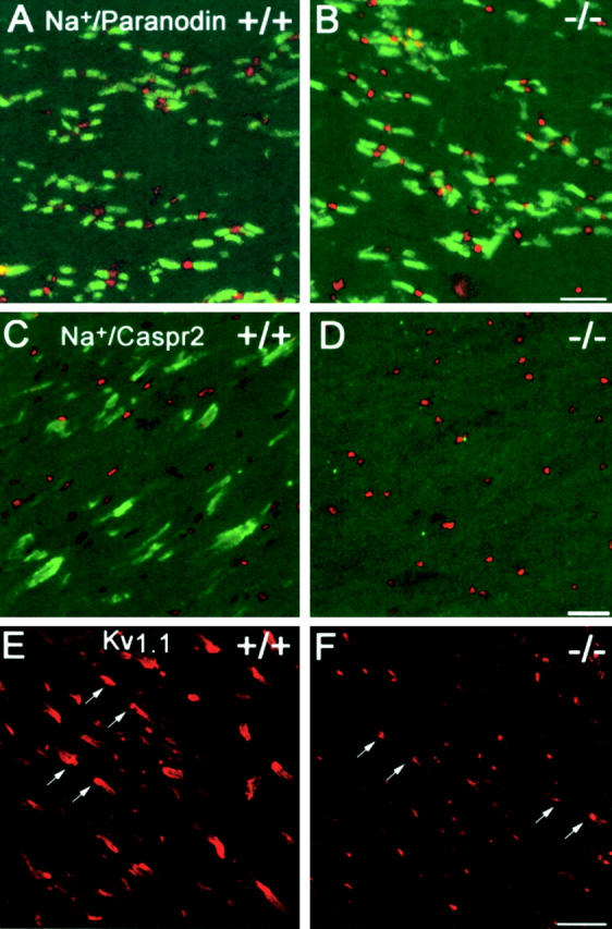
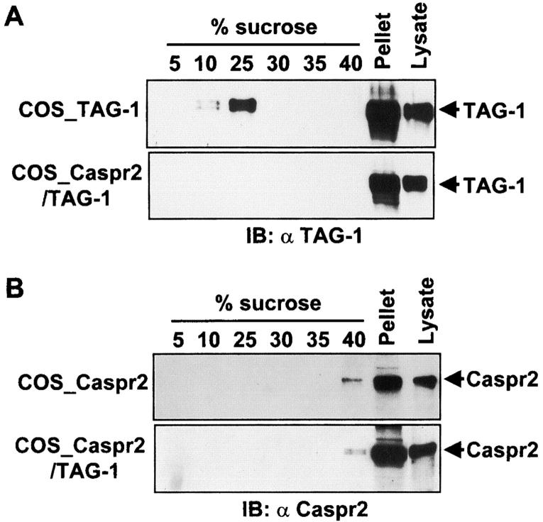
Myelination results in a highly segregated distribution of axonal membrane proteins at nodes of Ranvier. Here, we show the role in this process of TAG-1, a glycosyl-phosphatidyl-inositol-anchored cell adhesion molecule. In the absence of TAG-1, axonal Caspr2 did not accumulate at juxtaparanodes, and the normal enrichment of shaker-type K+ channels in these regions was severely disrupted, in the central and peripheral nervous systems. In contrast, the localization of protein 4.1B, an axoplasmic partner of Caspr2, was only moderately altered. TAG-1, which is expressed in both neurons and glia, was able to associate in cis with Caspr2 and in trans with itself. Thus, a tripartite intercellular protein complex, comprised of these two proteins, appears critical for axo-glial contacts at juxtaparanodes. This complex is analogous to that described previously at paranodes, suggesting that similar molecules are crucial for different types of axo-glial interactions.


Figures

Figure 1.
Figure 1.
Ultrastructural organization of myelinated fibers and nerve function in TAG-1 mutant mice. (A–F) Ultrastructural organization of myelinated fibers. Myelin sheath thickness and compaction were similar in myelinated fibers of the ventral spinal cord region of 3-mo-old (A) wild-type and (B) TAG-1 mutant mice. The nodal (N), paranodal (P), and juxtaparanodal (J) regions appeared properly organized in longitudinal sections of wild-type (C) and mutant animals (D). In the paranodal region, the transverse bands (arrowheads) were normally present in both genotypes (E, wild type; F, mutant). Bars: (A and B) 0.1 μm; (C and D) 0.5 μm; and (E and F) 0.3 μm. (G and H) Electrophysiological studies of sciatic nerves. CMAPs and F waves were recorded after distal and proximal stimulation of sciatic nerves of 2-mo-old (G) wild-type and (H) TAG-1 mutant mice. There were no differences in the waveform, latencies and amplitudes of CMAPs between both genotypes.
Figure 2.
Figure 2.
Distribution of specific proteins in myelinated optic nerves of TAG-1 mutant mice. Localization of molecular components of nodes, paranodes, and juxtaparanodes in optic nerve sections of 2-mo-old (A, C, and E) wild-type (+/+) and (B, D, and F) TAG-1 mutant (−/−) mice. Sodium channels (red) and paranodin/Caspr (green) were normally clustered in the nodal and paranodal regions, respectively, in both (A) wild-type and (B) TAG-1–deficient mice. Caspr2-IR (green) was normally detected in the juxtaparanodal regions, in reference to the nodal sodium channels (red) in (C) wild-type animals, whereas it was not visible in (D) TAG-1 mutant mice. Kv1.1-IR was dramatically altered in mutant mice (F, arrows), as compared with wild-type animals (E, arrows). In TAG-1 mutant mice, Kv1.1 labeling was markedly decreased and was mostly restricted to a small area in contact with (F) paranodes. Bars: (A–F) 5 μm.
Figure 3.
Figure 3.
Expression levels of juxtaparanodal proteins in central myelinated fibers of TAG-1 knockout mice. Expression levels of (A) TAG-1, (B) Caspr2, and (C) Kv1.1 potassium channels in 2-mo-old wild-type (+/+) and TAG-1 mutant mice (−/−) were examined by IB analysis of optic nerve extracts (top). Protein levels were quantified (three mice in each group) using actin (bottom) in each sample for normalization: Caspr2 levels in mutant mice were 84 ± 2% of wild-type (mean ± SD); Kv1.1 86-kD band; 129± 22%; and 70-kD band 193 ± 21%. The position of molecular mass markers (kD) is indicated.
Figure 4.
Figure 4.
Distribution of specific proteins in sciatic nerves nodal regions of TAG-1 mutants and during development. (A–L) Localization of molecular components of nodes, paranodes, and juxtaparanodes in teased sciatic nerve fibers of 2-mo-old (A, C, E, G, I, and K) wild-type (+/+) and (B, D, F, H, J, and L) TAG-1 mutant (−/−) mice: (A and B) TAG-1 (red) and paranodin/Caspr (green); (C and D) NF155; (E and F) sodium channels; (G and H) MAG (green, paranodal regions, arrows; Schmidt-Lanterman incisures, arrowheads) and Caspr2 (red); (I and J) Kv1.1 potassium channels (green) and Caspr/paranodin (red); (K and L) Protein 4.1B (red) and Caspr/paranodin (green). Caspr2 and Kv1.1 labeling were dramatically altered in mutant mice, whereas the other markers appeared normal. Protein 4.1B-IR (red) was intense in the paranodal regions (arrows) where it was colocalized with Caspr/paranodin (green), and was also visible in the juxtaparanodal regions (arrowheads) of wild-type and mutant mice. Bars: (A–J) 20 μm and (K and L) 5 μm. (M and N) Distribution of Caspr2, TAG-1 and Kv1.2 in teased fibers of sciatic nerve of a P8 rat. (M) Caspr2 (red) and (N) TAG-1 (red) were detected in a limited number of fibers, where they appeared colocalized with (M and N) Kv1.2 potassium channels (green). Bar, 2.5 μm.
Figure 5.
Figure 5.
Association of TAG-1 and Caspr2 in brain and transfected COS-7 cells. (A and B) Association of TAG-1 and Caspr2 in brain. Rat brain proteins were extracted and subjected to IP with (A) αCaspr2 or (B) αTAG-1. The presence of specific proteins in the precipitates was examined by IB with the indicated antibodies. Aliquots of crude protein extracts (Lysate, 1/60 of protein amount used for each coIP) were also subjected to IB to verify the expression of the proteins. (A) Caspr2 was detected in immune precipitates with TAG-1 antibodies but not with antibodies against other IgSF proteins (αNrCAM, αL1, and αF3). (B) TAG-1 was detected in immune precipitates with Caspr2 antibodies but not nonimmune serum (αNI). (C and D) Association of TAG-1 and Caspr2 in transfected COS-7 cells. Lysates from COS-7 cells overexpressing either Caspr2 or TAG-1 alone, or both, were prepared as described in Materials and methods and subjected to IP either with (C) αTAG-1 or (D) αCaspr2. Precipitates were resolved by SDS-PAGE, analyzed by IBs with antibodies against Caspr2 and TAG-1, to detect the presence of specific proteins (C and D, top and middle). Aliquots of crude protein extracts (Lysates) were also subjected to immunoblotting to verify the expression of the proteins (C and D, lower panels). TAG-1 antibodies coIP Caspr2 only in doubly transfected cells (C), whereas Caspr2 antibodies pulled-down TAG-1 only in cotransfected cells (D). Note the slight shift of migration of Caspr2 in the presence of TAG-1, which results in a doublet with a predominant lower band in cotransfected cells (C, Lysates). The position of molecular mass markers (kD) is indicated.
Figure 6.
Figure 6.
Cis and trans interactions between TAG-1 and Caspr2 in COS-7 cells. (A–C) Colocalization of Caspr2 and TAG-1 at the plasma membrane of COS-7 cells. COS-7 cells overexpressing either Caspr2 or TAG-1 alone, or both, were processed for indirect IF and laser confocal microscopy analysis. Caspr2 was uniformly localized at the plasma membrane (A, COS_Caspr2). TAG-1 displayed a membrane localization with a patchy appearance (B, COS_TAG-1). In cotransfected cells, Caspr2 (green) and TAG-1 (red) were largely colocalized (yellow) at the plasma membrane with a distribution similar to that of Caspr2 alone (C, COS_Caspr2/TAG-1). Single confocal sections are shown. (D) Association of TAG-1 in trans with itself but not with Caspr2. COS-7 cells overexpressing either TAG-1 alone (COS_TAG-1, a and b), Caspr2 (COS_Caspr2, c and d) or both (COS_Caspr2/TAG-1, e and f) were incubated with a (a and c–f) TAG-1-Fc chimeric protein or a (b) MUC-18-Fc chimera protein, and processed for indirect IF and laser confocal microscopy analysis. The TAG-1-Fc chimeric protein (green) bound to cells expressing (a) TAG-1 alone or in combination with (e) Caspr2 but not to cells expressing (c) Caspr2 alone. Expression of Caspr2 was verified by (d and f) specific IF (red). Stacked confocal images of eight consecutive sections 1 μm apart. Bar, (A–D) 10 μm.
Figure 7.
Figure 7.
Sucrose density gradient of TAG-1 and Caspr2 in transfected COS-7 cells. COS-7 cells overexpressing either (A, top, COS_TAG-1) TAG-1 alone or (B, top, COS_Caspr2) Caspr2 alone, or (A and B, bottom, COS_Caspr2/TAG-1) both proteins, were lysed in a Triton X-100 containing buffer and lysates were submitted to discontinuous sucrose gradients as described in Materials and methods and IB with antibodies against (A) TAG-1 and (B) Caspr2. Aliquots of crude protein extracts (Lysate) were also analyzed to verify the expression of the proteins. TAG-1 expressed alone was present in the 10 and 25% sucrose fractions (A, top). In contrast, Caspr2 was recovered in the heavier 40% fraction and in the pellet (B, bottom). When coexpressed with Caspr2, TAG-1 was no longer present in the light fractions but was found in the pellet with Caspr2 (A and B, bottom).
Figure 8.
Figure 8.
Model of the molecular organization of juxtaparanodal regions. This model is the simplest that can account for the data from previous studies and the present work. Caspr2 is enriched in the axolemma, whereas TAG-1 is expressed in both neurons and myelinating glial cells. Experiments in transfected cells show that TAG-1 interacts with Caspr2 in cis, and that TAG-1 exchanges trans interactions with itself (homophilic) but not with Caspr2. The functional importance of these interactions is demonstrated by the absence of Caspr2 enrichment in juxtaparanodal regions in TAG-1 knockout mice. These complexes are associated with other proteins including Kv1.1 and Kv1.2 potassium channels, presumably through a PDZ domain–containing protein (schematically represented here; Poliak et al., 1999; Rasband et al., 2002) and with protein 4.1B (Denisenko-Nehrbass et al., 2003b). Because protein 4.1B was still detected at juxtaparanodes in the absence of TAG-1 and Caspr2, it is likely that it interacts with other proteins, including components of the cytoskeleton (arrow). Correct localization of juxtaparanodal proteins may depend on both axo–glial interactions and binding to axoplasmic cytoskeletal components.

Similar articles

Cited by

References

    1. Arroyo, E.J., T. Xu, J. Grinspan, S. Lambert, S.R. Levinson, P.J. Brophy, E. Peles, and S.S. Scherer. 2002. Genetic dysmyelination alters the molecular architecture of the nodal region. J. Neurosci. 22:1726–1737. - PMC - PubMed
    1. Baumgartner, S., J.T. Littleton, K. Broadie, M.A. Bhat, R. Harbecke, J.A. Lengyel, R. Chiquet-Ehrisman, A. Prokop, and H.J. Bellen. 1996. A Drosophila neurexin is required for septate junction and blood-nerve barrier formation and function. Cell. 87:1059–1068. - PubMed
    1. Bhat, M.A., J.C. Rios, Y. Lu, G.P. Garci-Fresco, W. Ching, M. St Martin, J. Li, and S. Einheber. 2001. Axon-glia interactions and the domain organization of myelinated axons requires neurexin IV/Caspr/Paranodin. Neuron. 30:369–383. - PubMed
    1. Boyle, M.E., E.O. Berglund, K.K. Murai, L. Weber, E. Peles, and B. Ranscht. 2001. Contactin orchestrates assembly of the septate-like junctions at the paranode in myelinated peripheral nerve. Neuron. 30:385–397. - PubMed
    1. Buttiglione, M., J.M. Revest, O. Pavlou, D. Karagogeos, A. Furley, G. Rougon, and C. Faivre-Sarrailh. 1998. A functional interaction between the neuronal adhesion molecules TAG-1 and F3 modulates neurite outgrowth and fasciculation of cerebellar granule cells. J. Neurosci. 18:6853–6870. - PMC - PubMed

Publication types

MeSH terms