

Three dimensional volume quantification of aortic valve calcification using multislice computed tomography
- PMID: 12975416
- PMCID: PMC1767906
- DOI: 10.1136/heart.89.10.1191
Three dimensional volume quantification of aortic valve calcification using multislice computed tomography
Abstract
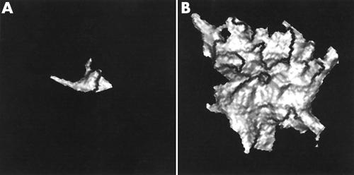
Objective: To assess a new multislice computed tomography (CT) technique for three dimensional quantification of aortic valve calcification volume (3D AVCV) and to study the relation between stenosis and calcification of the aortic valve.
Methods: 50 patients with echocardiographic calcification of the aortic valve underwent two separate ECG triggered multislice CT for quantification of 3D AVCV. The agreement between the two 3D AVCV scores was assessed and 3D AVCV was compared with echocardiographic markers of severity of aortic stenosis.
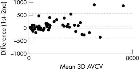
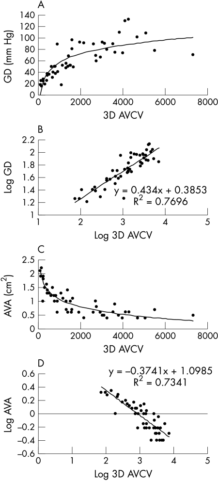
Results: Overall the level of agreement between the two 3D AVCV scores was excellent (median interscan variability 7.9% (interquartile range 10.1); correlation coefficient, r = 0.99; repeatability coefficient 237.8 mm3 (limits of agreement -393 to 559 mm3)). However, the magnitude of the 3D AVCV did influence the interscan variability. The 3D AVCV correlated closely with the maximal predicted transvalvar gradient (r2 = 0.77) and aortic valve area (r2 = 0.73).
Conclusions: Multislice CT provides a technique for quantifying 3D AVCV that has good reproducibility. There is a close non-linear relation between echocardiographic parameters of severity of valve stenosis and 3D AVCV scores.
Figures
Similar articles
-
Preliminary experience in the assessment of aortic valve calcification by ECG-gated multislice spiral computed tomography.Int J Cardiol. 2005 Jul 10;102(2):195-200. doi: 10.1016/j.ijcard.2004.05.017. Int J Cardiol. 2005. PMID: 15982484
-
Aortic valve calcification on computed tomography predicts the severity of aortic stenosis.Clin Radiol. 2003 Sep;58(9):712-6. doi: 10.1016/s0009-9260(03)00184-3. Clin Radiol. 2003. PMID: 12943644
-
Electrocardiographically gated multi-detector row CT for assessment of valvular morphology and calcification in aortic stenosis.Radiology. 2002 Oct;225(1):120-8. doi: 10.1148/radiol.2251011703. Radiology. 2002. PMID: 12354994
-
Role of preprocedural computed tomography in transcatheter aortic valve implantation.Rofo. 2013 Oct;185(10):941-9. Rofo. 2013. PMID: 24490256 Review.
-
Aortic valve imaging with computed tomography: a review.J Heart Valve Dis. 2002 Sep;11(5):604-11. J Heart Valve Dis. 2002. PMID: 12358394 Review.
Cited by
-
Role of Computational Simulations in Heart Valve Dynamics and Design of Valvular Prostheses.Cardiovasc Eng Technol. 2010 Mar;1(1):18-38. doi: 10.1007/s13239-010-0002-x. Cardiovasc Eng Technol. 2010. PMID: 20606715 Free PMC article.
-
CT of valvular heart disease.Int J Cardiovasc Imaging. 2005 Feb;21(1):105-13. doi: 10.1007/s10554-004-5339-5. Int J Cardiovasc Imaging. 2005. PMID: 15915944 Review.
-
Semiautomatic, Quantitative Measurement of Aortic Valve Area Using CTA: Validation and Comparison with Transthoracic Echocardiography.Biomed Res Int. 2015;2015:648283. doi: 10.1155/2015/648283. Epub 2015 Jun 29. Biomed Res Int. 2015. PMID: 26221603 Free PMC article.
-
Pictorial essay: Non-coronary applications of cardiac CT.Indian J Radiol Imaging. 2012 Jan;22(1):40-6. doi: 10.4103/0971-3026.95403. Indian J Radiol Imaging. 2012. PMID: 22623815 Free PMC article.
-
Cardiac CT: coronary arteries and beyond.Eur Radiol. 2007 Apr;17(4):994-1008. doi: 10.1007/s00330-006-0433-9. Epub 2006 Oct 26. Eur Radiol. 2007. PMID: 17066290 Review.
References
-
- Rosenhek R, Binder T, Porenta G, et al. Predictors of outcome in severe, asymptomatic aortic stenosis. N Engl J Med 2000;343:611–7. - PubMed
-
- Pohle K, Mäffert R, Ropers D, et al. Progression of aortic valve calcification associated with coronary atherosclerosis and cardiovascular risk factors. Circulation 2001;104:1927–32. - PubMed
-
- Shavelle D, Takasu J, Budoff M. HMG CoA reductase inhibitor (statin) and aortic valve calcium. Lancet 2002;359:1125–6. - PubMed
-
- Melina G, Rubens M, Amrani M, et al. Electron beam tomography for cusp calcification in homograft versus freestyle xenografts. Ann Thorac Surg 2001;71:S368–70. - PubMed
-
- Ohnesorge B, Flohr T, Fischbach R, et al. Reproducibility of coronary calcium quantification in repeat examinations with retrospectively ECG-gated multisection spiral CT. Eur Radiol 2002;12:1532–40. - PubMed
Publication types
MeSH terms
LinkOut - more resources
Full Text Sources
Other Literature Sources
Medical