. 2003 Oct;89(10):1211-6.
doi: 10.1136/heart.89.10.1211.

Management of fetal tachyarrhythmia based on superior vena cava/aorta Doppler flow recordings

Affiliations

Management of fetal tachyarrhythmia based on superior vena cava/aorta Doppler flow recordings

J-C Fouron et al. Heart. 2003 Oct.

Abstract

Objective: To evaluate a management protocol of fetal supraventricular tachycardia (SVT) based on prior identification of the underlying mechanism.

Design and setting: Prospective study in a mother-child tertiary university centre.

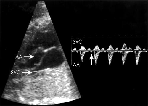
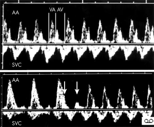
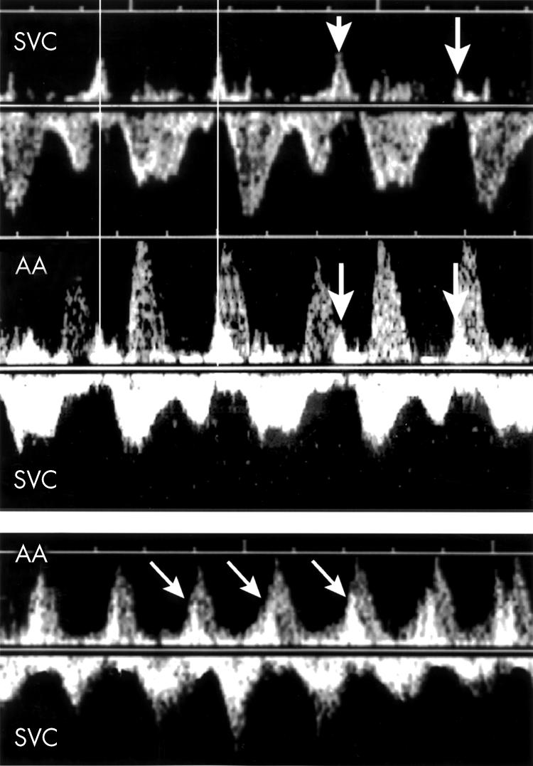
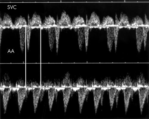
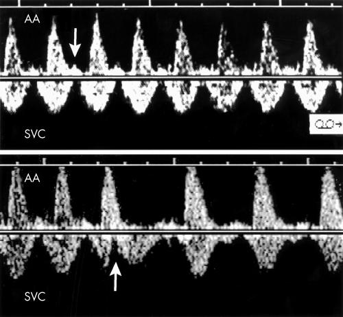
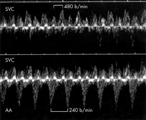
Patients: During a consecutive 36 month period, 18 fetuses with sustained SVT underwent a superior vena cava/ascending aorta (SVC/AA) Doppler investigation in an attempt to determine the atrioventricular (AV) relation and to treat the arrhythmia according to a pre-established management protocol.

Main outcome measure: Rate of conversion to sinus rhythm.

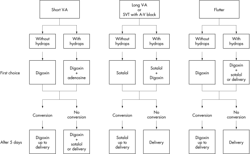
Results: Seven fetuses had short ventriculoatrial tachycardia, five of these with a 1:1 AV conduction suggesting re-entrant tachycardia. The first choice drug was digoxin and all were converted. One fetus had AV dissociation leading to the diagnosis of junctional ectopic tachycardia, which was resistant to digoxin and sotalol; amiodarone achieved postnatal conversion. One fetus had SVT and first or second AV block; the diagnosis was atrial ectopic tachycardia (AET), which responded to sotalol given as a drug of first choice. Seven fetuses had long ventriculoatrial tachycardia: one with sinus tachycardia (no treatment), one with permanent junctional reciprocating tachycardia (PJRT), and three with AET. The first choice drug was sotalol and all were converted. One AET was classified postnatally as PJRT. Six fetuses had intra-atrial re-entrant tachycardia: five with 2:1 AV conduction and one with variable block. The first choice drug was digoxin. Conversion was achieved in all but one, who died after birth from advanced cardiomyopathy.

Conclusion: The electrophysiological mechanisms of fetal SVT can be clarified with SVC/AA Doppler. The proposed management protocol has so far yielded a good rate of conversion to sinus rhythm.


Figures

Figure 1
Figure 1
Left: Two dimensional imaging of a segment of the ascending aorta (AA) adjacent to the superior vena cava (SVC) draining into the right atrium. The widened Doppler sampling volume encompasses the aorta and SVC. Right: Doppler tracing obtained from the same patient. Aortic ejection waves are recorded below the zero velocity line. Anterograde venous flow in the SVC going in the opposite direction is above the same line. Each aortic ejection wave is preceded by a small venous retrograde wave caused by right atrial contraction (arrow).
Figure 2
Figure 2
Treatment protocol for fetal tachyarrhythmia. AV, atrioventricular; SVT, supraventricular tachycardia; VA, ventriculoatrial.
Figure 3
Figure 3
Top: SVC/AA recording in a case (fetus 2) of atrioventricular re-entrant tachycardia. Bottom: In the same patient, sudden onset of the tachycardia triggered by supraventricular couplets (arrows) is shown.
Figure 4
Figure 4
Top: Doppler flow velocity recordings in the SVC of fetus 5 showing an atrial rate of 130 beats/min (arrows). Middle: The SVC/AA recording shows that the ventricles are contracting at the faster rate of 200 beats/min leading to the diagnosis of ventricular tachycardia with AV dissociation. The two vertical lines join the venous “a” waves and illustrate that the atrial rate of contraction is the same on both tracings. Bottom: During a brief period of 1:1 conduction in the same patient, the SVC/AA Doppler tracing shows atrial contractions (arrows) occurring simultaneously or a few milliseconds after ventricular contractions, confirming the diagnosis of junctional ectopic tachycardia.
Figure 5
Figure 5
Pulsed Doppler tracing obtained from fetus 6. Top: A fixed atrial rate at 220 beats/min is associated with a 2:1 atrioventricular conduction. Bottom: The atrial rate is the same (vertical lines) but a 1:1 conduction is observed. The AV time interval is then so prolonged that the VA appears shorter than the AV. In 1:1 conduction, the A waves are prominent because atrial contractions occur while the tricuspid valve is closed. In the presence of a 2:1 relation (top), the blocked A waves are even taller due to greater blood volume accumulated in the right atrium.
Figure 6
Figure 6
Top: Example of a long VA tachycardia. The A waves are of normal amplitudes (arrow). Bottom: In the same patient, sudden arrest of the tachycardia is observed following premature atrial contraction (arrow), suggesting the possibility of a permanent junctional reciprocating tachycardia.
Figure 7
Figure 7
Top: Doppler flow velocity waveforms in the SVC of fetus 15 showing atrial flutter waves at the rate of 480 beats/min. Bottom: The SVC/AA recording in the same patient shows a 2:1 relation between the flutter waves and ventricular contractions.

Similar articles

Cited by

References

    1. Benditt DG, Pritchett ELC, Smith WM, et al. Ventriculoatrial intervals: diagnostic use in paroxysmal supraventricular tachycardia. Ann Intern Med 1979;91:161–6. - PubMed
    1. Jaeggi E, Fouron JC, Fournier A, et al. Ventriculo-atrial time interval measured on M-mode echocardiography: a determining element in diagnosis, treatment, and prognosis of fetal supraventricular tachycardia. Heart 1998;79:582–7. - PMC - PubMed
    1. Fouron JC, Proulx F, Miró J, et al. Doppler and M-mode ultrasonography to time fetal atrial and ventricular contractions. Obstet Gynecol 2000;96:732–6. - PubMed
    1. Erkkola R, Lammintausta R, Liukko P, et al. Transfer of propanolol and sotalol across the human placenta: their effect on maternal and fetal plasma renin activity. Acta Obstet Gynecol Scand 1982;61:31–4. - PubMed
    1. Houyel L, Fournier A, Ducharme G, et al. Electrophysiologic effects of sotalol on the immature mammalian heart. J Cardiovasc Pharmacol 1992;19:134–9. - PubMed

MeSH terms

Substances