. 2003 Sep 15;198(6):889-901.
doi: 10.1084/jem.20030171.

CD4+CD25+ T cells regulate virus-specific primary and memory CD8+ T cell responses

Affiliations

CD4+CD25+ T cells regulate virus-specific primary and memory CD8+ T cell responses

Susmit Suvas et al. J Exp Med. .

Abstract

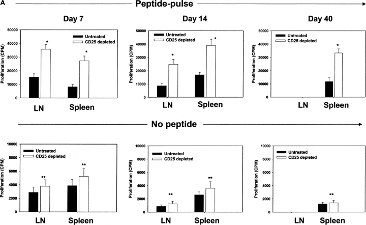
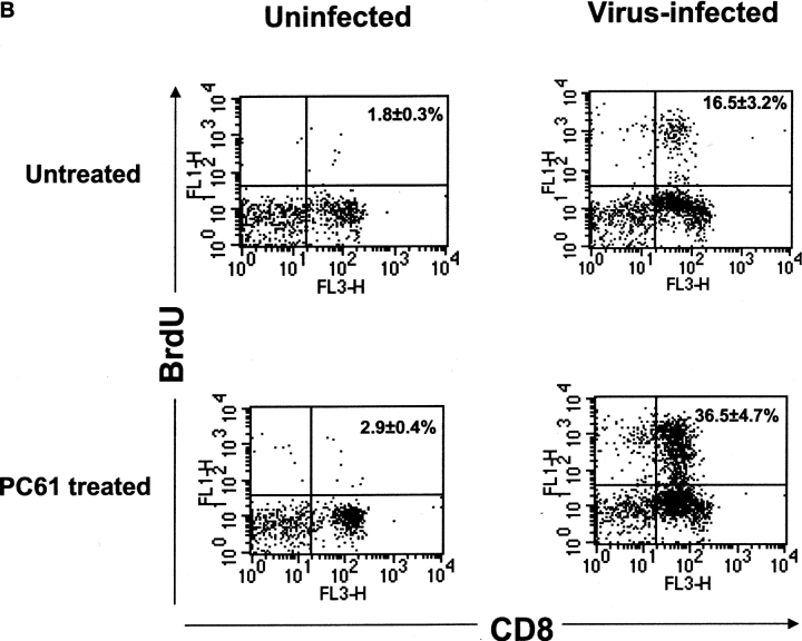
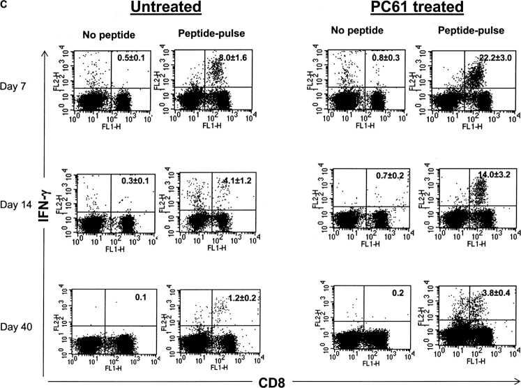
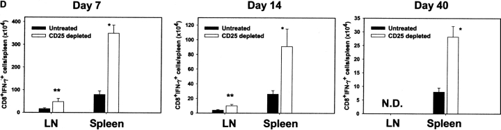
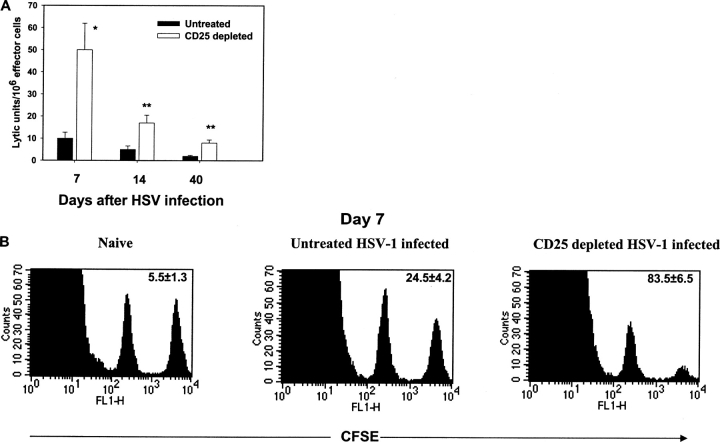
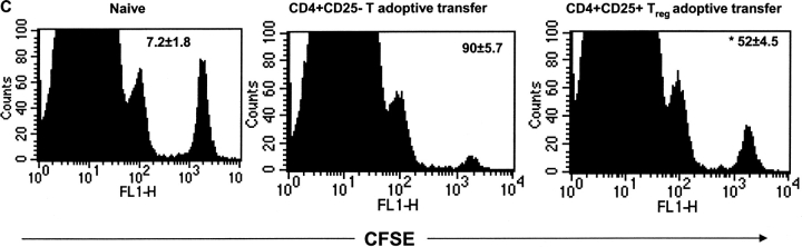
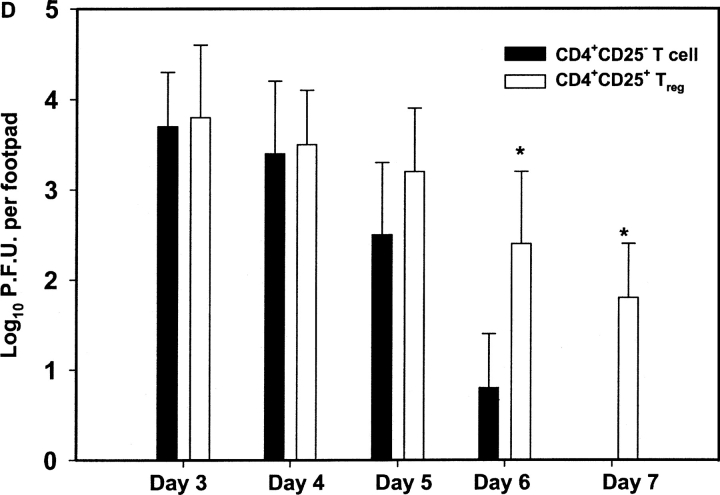
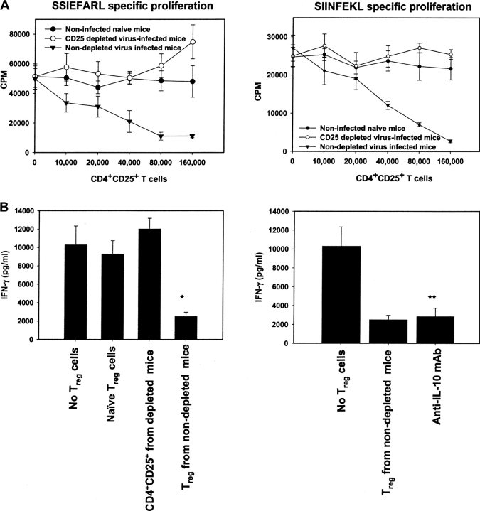
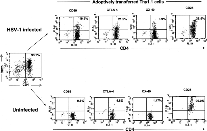
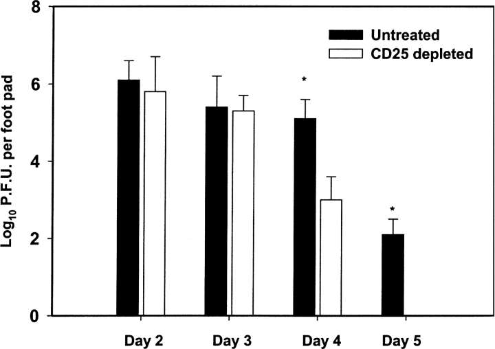
Naturally occurring CD4+CD25+ regulatory T cells appear important to prevent activation of autoreactive T cells. This article demonstrates that the magnitude of a CD8+ T cell-mediated immune response to an acute viral infection is also subject to control by CD4+CD25+ T regulatory cells (Treg). Accordingly, if natural Treg were depleted with specific anti-CD25 antibody before infection with HSV, the resultant CD8+ T cell response to the immunodominant peptide SSIEFARL was significantly enhanced. This was shown by several in vitro measures of CD8+ T cell reactivity and by assays that directly determine CD8+ T cell function, such as proliferation and cytotoxicity in vivo. The enhanced responsiveness in CD25-depleted animals was between three- and fourfold with the effect evident both in the acute and memory phases of the immune response. Surprisingly, HSV infection resulted in enhanced Treg function with such cells able to suppress CD8+ T cell responses to both viral and unrelated antigens. Our results are discussed both in term of how viral infection might temporarily diminish immunity to other infectious agents and their application to vaccines. Thus, controlling suppressor effects at the time of vaccination could result in more effective immunity.


Figures

Figure 1.
Figure 1.
Enhancement of antigen-specific CD8+ T cell responses after in vivo depletion of CD4+CD25+ Treg cells before HSV-1 infection. (A) The kinetics of SSIEFARL-specific CD8+ T cell proliferation was measured in vitro in both depleted and nondepleted mice by [3H]thymidine incorporation. The results are expressed as cpm. *P < 0.01 compared with nondepleted HSV-infected B6 mice. (N.D., not done). The experiment shown is representative of two similar experiments, and error bars reflect the mean ± SD of four mice per group. The cpm of nondepleted and CD25+-depleted noninfected groups was always <1,000 with no significant difference observed between the two groups. (B) In vivo proliferation of Vβ10+CD8+ T cells was measured by BrdU incorporation assay. Mice infected with HSV-1 3 d postdepletion were injected i.p. with 100 μl (1 mg) of BrdU solution in PBS 1 d before sacrifice. Mice were terminated 7 d p.i. to examine the in vivo proliferation of CD8+Vβ10+T cells. Cells were gated on Vβ10+ cells, and the percentage of BrdU+CD8+Vβ10+ T cells is represented in the upper right quadrant. The data in B are representative of two identical experiments. (C) Kinetics of HSV-1–specific immune response shows that SSIEFARL-specific CD8+ T cells increased in number in PC61-treated mice are also functional as evident by intracellular IFN-γ staining. The experiment shown is representative of two additional experiments. The values shown in these FACS® plots are the mean of four mice per group. (D) Increase in IFN-γ–secreting CD8+ T cells in B6 mice devoid of CD4+CD25+ T cells were also measured by standard ELISPOT assay. On different days post HSV infection, LNs and spleen cells were analyzed for the number of IFN-γ–secreting CD8+ T cells in response to SSIEFARL peptide. The bars represent the mean ± SD of three different mice of same group and are representative of two similar experiments. *P < 0.01 and **P < 0.05 compared with untreated HSV-infected B6 mice. (N.D., not done). Without peptide stimulation, there was no significant difference in CD8/IFN-γ–producing cells in nondepleted and CD25-depleted HSV-infected mice, and the maximum number obtained at any time point was always <5,000 per spleen.
Figure 1.
Figure 1.
Enhancement of antigen-specific CD8+ T cell responses after in vivo depletion of CD4+CD25+ Treg cells before HSV-1 infection. (A) The kinetics of SSIEFARL-specific CD8+ T cell proliferation was measured in vitro in both depleted and nondepleted mice by [3H]thymidine incorporation. The results are expressed as cpm. *P < 0.01 compared with nondepleted HSV-infected B6 mice. (N.D., not done). The experiment shown is representative of two similar experiments, and error bars reflect the mean ± SD of four mice per group. The cpm of nondepleted and CD25+-depleted noninfected groups was always <1,000 with no significant difference observed between the two groups. (B) In vivo proliferation of Vβ10+CD8+ T cells was measured by BrdU incorporation assay. Mice infected with HSV-1 3 d postdepletion were injected i.p. with 100 μl (1 mg) of BrdU solution in PBS 1 d before sacrifice. Mice were terminated 7 d p.i. to examine the in vivo proliferation of CD8+Vβ10+T cells. Cells were gated on Vβ10+ cells, and the percentage of BrdU+CD8+Vβ10+ T cells is represented in the upper right quadrant. The data in B are representative of two identical experiments. (C) Kinetics of HSV-1–specific immune response shows that SSIEFARL-specific CD8+ T cells increased in number in PC61-treated mice are also functional as evident by intracellular IFN-γ staining. The experiment shown is representative of two additional experiments. The values shown in these FACS® plots are the mean of four mice per group. (D) Increase in IFN-γ–secreting CD8+ T cells in B6 mice devoid of CD4+CD25+ T cells were also measured by standard ELISPOT assay. On different days post HSV infection, LNs and spleen cells were analyzed for the number of IFN-γ–secreting CD8+ T cells in response to SSIEFARL peptide. The bars represent the mean ± SD of three different mice of same group and are representative of two similar experiments. *P < 0.01 and **P < 0.05 compared with untreated HSV-infected B6 mice. (N.D., not done). Without peptide stimulation, there was no significant difference in CD8/IFN-γ–producing cells in nondepleted and CD25-depleted HSV-infected mice, and the maximum number obtained at any time point was always <5,000 per spleen.
Figure 2.
Figure 2.
Increased antigen-specific CD8+ T cell cytotoxicity in CD25-depleted B6 mice after HSV-1 infection. (A) Splenocytes collected from WT and CD25-depleted B6 mice at different time points after HSV infection were expanded in vitro with irradiated SSIEFARL-pulsed syngeneic splenocytes for 5 d and used as effectors in a 4-h 51Cr release assay. The targets included SSIEFARL-pulsed, MHC-matched MC-38 and unpulsed MC-38. The figure shows only the data for SSIEFARL peptide-pulsed MC-38 targets in lytic units. One lytic unit (LU) is the number of lymphocytes required to give 30% lysis. The lysis in control targets was insignificant and below the limit for calculation of lytic units. The experiments were performed using four mice, and data shown above are mean ± SD of four mice from one experiment. *P < 0.01 and **P < 0.05 compared with untreated HSV-infected B6 mice. (B) To analyze the difference of in vivo SSIEFARL-specific cytotoxicity in vivo in CD25-depleted and nondepleted B6 mice 7 d post HSV-1 infection, syngeneic spleen cells were pulsed with gB498–505 peptide and labeled with CFSE (2.5 μm). To control for antigen specificity, unpulsed syngeneic spleen cells were labeled with a lower concentration of CFSE (0.25 μm). A 1:1 mixture of each target cell population was injected i.v. into both groups of mice. 1 h p.i., spleen cells were prepared from individual mice and acquired on a FACS® Vantage system. The percentage of specific lysis was determined as mentioned in Materials and Methods. The number shown in each plot is the mean of percent antigen specific lysis observed in four mice per group.
Figure 3.
Figure 3.
Increased rate of viral clearance from the FP of CD25-depleted HSV-1–infected mice during the primary immune response. Mice were infected in the hind FPs with 106 pfu HSV-1, and on days 3 to 6 the hind feet were removed and homogenized in virus diluent. The resulting supernatant was used in a vero cell based viral plaque assay to determine the virus titer. The results are expressed as number of pfu of virus per FP. Each error bar represents the mean and SD of six individual FPs. *P < 0.01 compared with CD25-depleted HSV-infected B6 mice.
Figure 4.
Figure 4.
Removal of CD4+CD25+ T cells before HSV infection enhances the expression of activation markers on virus-specific CD8+ T cells. Expression of activation markers on T cells gated on with double positive CD8+Vβ10+ cells in spleen of CD25-depleted and nondepleted mice were examined. Splenocytes from both groups were collected on day 14 post HSV infection and stained with anti–mouse CD8α and anti–mouse Vβ10 mAb in combination with an antibody against on activation marker (anti-CD44, CD62L, CD69, CD25, CCR5) and the appropriate isotype control using three color FACS® analysis. Histograms are representative of four individual mice from one such experiment, and values reflect the percentage of cells expressing the respective marker.
Figure 5.
Figure 5.
Adoptive transfer of CD4+CD25+ T cells inhibits HSV-1–specific CD8+ T cell responses. Purified CD4+CD25+ and CD4+CD25 T cells (2 × 10⁶/mouse) were adoptively transferred into WT B6 mice 24 h before HSV infection, and the immune response was measured on days 7 and 28 p.i. (A) On days 7 and 28 p.i., spleen cells were incubated with gB498–505, and CD8/IFN-γ production was measured by intracellular staining. The number shown in each plot is the mean percentage of IFN-γ–producing CD8+ T cells obtained from four mice per group. (B) The resulting decrease in IFN-γ–secreting CD8+ T cells in B6 mice after adoptive transfer of CD4+CD25+ T cells were also measured by a standard ELISPOT assay. On days 7 and 28 post HSV infection, spleen cells were analyzed for the number of IFN-γ–secreting CD8+ T cells in response to SSIEFARL peptide. The error bars represent the mean ± SD of four different mice in the same group. *P < 0.05 compared with HSV-infected B6 mice receiving no adoptive transfer. Without peptide stimulation, there was no significant difference in CD8/IFN-γ–producing cells between HSV-infected mice receiving either CD4+CD25+ or CD4+CD25 T cells. The maximum number of cells obtained at both time points was always <4,000 per spleen. (C) gB-specific cytotoxicity was also measured by an in vivo CTL assay to analyze differences in SSIEFARL-specific cytotoxicity after adoptive transfer of CD25+ or CD25 CD4+ T cells. On day 7 post HSV-1 infection, syngeneic spleen cells were pulsed with gB498–505 peptide and labeled with CFSE (2.5 μm). Unpulsed syngeneic spleen cells were labeled with a lower concentration of CFSE (0.25 μm). A 1:1 mixture of each target cell population was injected i.v. into both groups. 4 h p.i., spleen cells were prepared from individual mice and acquired on a FACS® vantage system. Specific lysis was calculated as mentioned in Materials and Methods. The number shown in each plot is the mean percent antigen specific lysis observed in four mice per group. *P < 0.05 compared with group with CD4+CD25 T cell adoptive transfer. (D) Adoptive transfer of CD25+ Treg delays the viral clearance in HSV-infected mice. Each bar represents mean and SD of six individual FPs. *P < 0.05 compared with HSV-infected B6 mice receiving CD4+CD25 T cells.
Figure 5.
Figure 5.
Adoptive transfer of CD4+CD25+ T cells inhibits HSV-1–specific CD8+ T cell responses. Purified CD4+CD25+ and CD4+CD25 T cells (2 × 10⁶/mouse) were adoptively transferred into WT B6 mice 24 h before HSV infection, and the immune response was measured on days 7 and 28 p.i. (A) On days 7 and 28 p.i., spleen cells were incubated with gB498–505, and CD8/IFN-γ production was measured by intracellular staining. The number shown in each plot is the mean percentage of IFN-γ–producing CD8+ T cells obtained from four mice per group. (B) The resulting decrease in IFN-γ–secreting CD8+ T cells in B6 mice after adoptive transfer of CD4+CD25+ T cells were also measured by a standard ELISPOT assay. On days 7 and 28 post HSV infection, spleen cells were analyzed for the number of IFN-γ–secreting CD8+ T cells in response to SSIEFARL peptide. The error bars represent the mean ± SD of four different mice in the same group. *P < 0.05 compared with HSV-infected B6 mice receiving no adoptive transfer. Without peptide stimulation, there was no significant difference in CD8/IFN-γ–producing cells between HSV-infected mice receiving either CD4+CD25+ or CD4+CD25 T cells. The maximum number of cells obtained at both time points was always <4,000 per spleen. (C) gB-specific cytotoxicity was also measured by an in vivo CTL assay to analyze differences in SSIEFARL-specific cytotoxicity after adoptive transfer of CD25+ or CD25 CD4+ T cells. On day 7 post HSV-1 infection, syngeneic spleen cells were pulsed with gB498–505 peptide and labeled with CFSE (2.5 μm). Unpulsed syngeneic spleen cells were labeled with a lower concentration of CFSE (0.25 μm). A 1:1 mixture of each target cell population was injected i.v. into both groups. 4 h p.i., spleen cells were prepared from individual mice and acquired on a FACS® vantage system. Specific lysis was calculated as mentioned in Materials and Methods. The number shown in each plot is the mean percent antigen specific lysis observed in four mice per group. *P < 0.05 compared with group with CD4+CD25 T cell adoptive transfer. (D) Adoptive transfer of CD25+ Treg delays the viral clearance in HSV-infected mice. Each bar represents mean and SD of six individual FPs. *P < 0.05 compared with HSV-infected B6 mice receiving CD4+CD25 T cells.
Figure 6.
Figure 6.
CD4+CD25+ T cells isolated from HSV-1–infected nondepleted B6 mice suppress CD8+ T cells proliferation to both HSV-specific and unrelated antigens. (A) HSV-1–primed CD8+ T cells or OT-1 CD8+ T cells (2 × 10⁶/ml) were stimulated with either SSIEFARL or SIINFEKL peptide-pulsed, T-depleted splenocytes (106/ml) in the presence of CD4+CD25+ T cells isolated from either naive, nondepleted, or CD25-depleted B6 mice 10 d post HSV-1 infection. The proliferation was measured in vitro by [3H]thymidine incorporation assay, and the results are expressed as cpm. The basal level of proliferation of CD4+CD25+ T cells isolated from each group was always >5,000 with maximum proliferation observed in CD4+CD25+ T cells purified from CD25-depleted virus-infected mice. (B) CD4+CD25+ T cells isolated from naive and virus-infected mice were cocultured (106/ml) with HSV-1–primed CD8+ T cells (2 × 10⁶/ml) stimulated with SSIEFARL-pulsed APCs (2 × 10⁶/ml), and IFN-γ secretion in cultures was measured by ELISA as mentioned in Materials and Methods. Anti–IL-10 mAb (5 μg/ml) was also added in some of the cocultures to determine the effect on IFN-γ production. No spontaneous IFN-γ production was observed in CD4+CD25+ T cells isolated from either group. *P < 0.01 when compared with wells containing no Treg cells and **P > 0.05 when compared with wells containing Treg. Results from one representative experiment are shown.
Figure 7.
Figure 7.
Activation of CD4+CD25+ T cells by HSV infection in an adoptive transfer model. CD4+CD25+ T cells purified from Thy1.1 B6 mice were adoptively transferred into congenic Thy1.2 B6 recipients. One group was infected 14 h posttransfer and the other acted as noninfected controls. On day 7, spleen cells isolated from both groups were gated on the Thy1.1 population and analyzed for the expression of the activation markers such as OX-40, CTLA-4, CD69, and CD25 on CD4+ T cells.
Figure 8.
Figure 8.
Adoptive transfer of HSV-specific memory CD8+ T cells generated in the absence of Treg results in enhanced viral clearance than those generated in the presence of CD25+ regulatory T cells. Splenic MACS purified CD8+ T cells isolated from depleted and nondepleted animals 90 d p.i. were adoptively transferred into CD25-depleted naive B6 mice. 24 h posttransfer, mice were challenged in the hind FPs with 5 × 10⁶ pfu of HSV-1 KOS. The level of infectious HSV in the FP was observed from day 2 to 5 postchallenge. *P < 0.05. A higher virus load was always observed in CD25-depleted, HSV-infected group without any adoptive transfer compared with the groups with adoptive transfer (data not depicted).

References

    1. Sakaguchi, S., N. Sakaguchi, M. Asano, M. Itoh, and M. Toda. 1995. Immunologic self-tolerance maintained by activated T cells expressing IL-2 receptor alpha-chains (CD25). Breakdown of a single mechanism of self-tolerance causes various autoimmune diseases. J. Immunol. 155:1151–1164. - PubMed
    1. Suri-Payer, E., A.Z. Amar, A.M. Thornton, and E.M. Shevach. 1998. CD4+CD25+ T cells inhibit both the induction and effector function of autoreactive T cells and represent a unique lineage of immunoregulatory cells. J. Immunol. 160:1212–1218. - PubMed
    1. Maloy, K.J., L. Salaun, R. Cahill, G. Dougan, N.J. Saunders, and F. Powrie. 2003. CD4+CD25+ T(R) cells suppress innate immune pathology through cytokine-dependent mechanisms. J. Exp. Med. 197:111–119. - PMC - PubMed
    1. McHugh, R.S., E.M. Shevach, and A.M. Thornton. 2001. Control of organ-specific autoimmunity by immunoregulatory CD4(+)CD25(+) T cells. Microbes Infect. 3:919–927. - PubMed
    1. Hori, S., T.L. Carvalho, and J. Demengeot. 2002. CD25+CD4+ regulatory T cells suppress CD4+ T cell-mediated pulmonary hyperinflammation driven by Pneumocystis carinii in immunodeficient mice. Eur. J. Immunol. 32:1282–1291. - PubMed

Publication types

MeSH terms