. 2003 Sep 15;198(6):903-11.
doi: 10.1084/jem.20022058.

Characterization of the CD4+ T cell response to Epstein-Barr virus during primary and persistent infection

Affiliations

Characterization of the CD4+ T cell response to Epstein-Barr virus during primary and persistent infection

Elisabeth Amyes et al. J Exp Med. .

Abstract

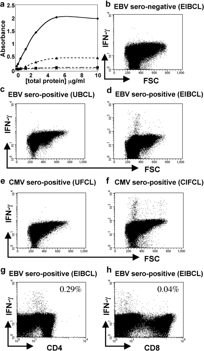
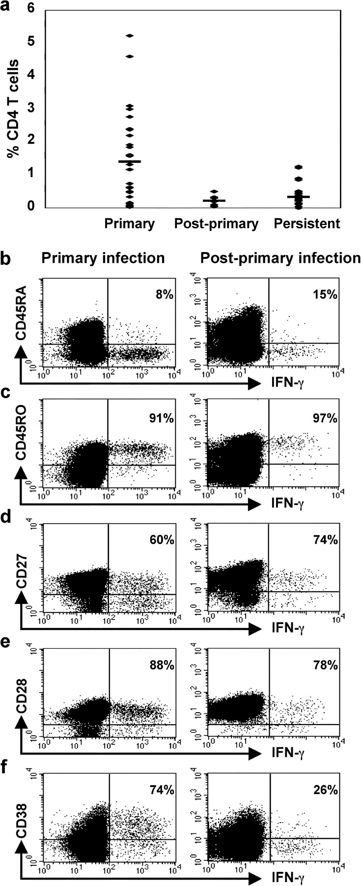
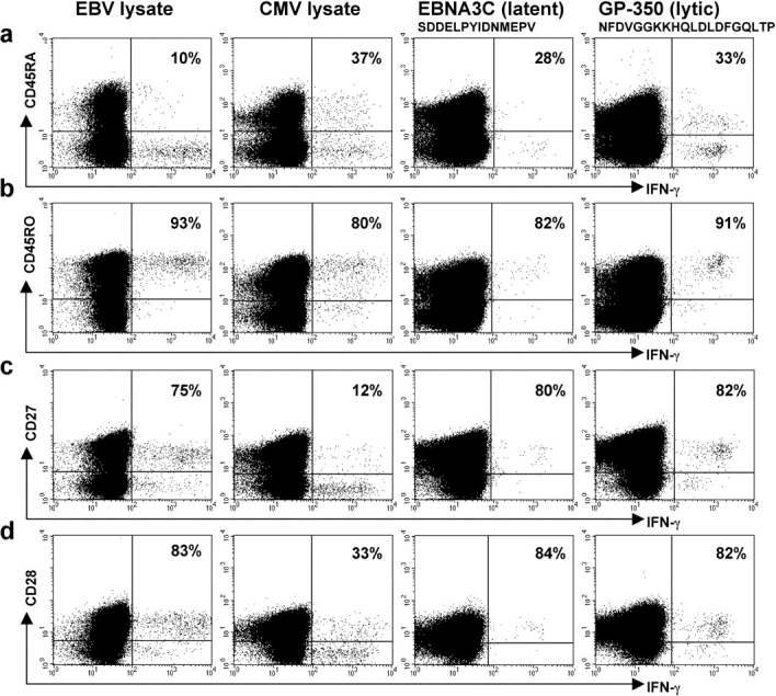
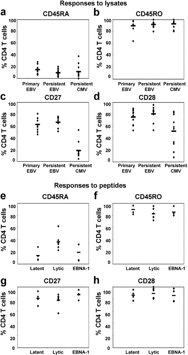
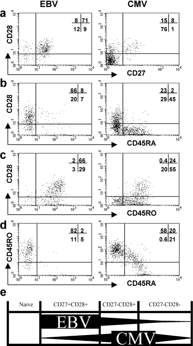
The CD8+ T cell response to Epstein-Barr virus (EBV) is well characterized. Much less is known about the evolution of the CD4+ T cell response. Here we show that EBV stimulates a primary burst of effector CD4+ T cells and this is followed by a period of down-regulation. A small population of EBV-specific effector CD4+ T cells survives during the lifelong persistent phase of infection. The EBV-specific effector CD4+ T cells accumulate within a CD27+ CD28+ differentiation compartment during primary infection and remain enriched within this compartment throughout the persistent phase of infection. Analysis of CD4+ T cell responses to individual epitopes from EBV latent and lytic cycle proteins confirms the observation that the majority of the effector cells express both CD27 and CD28, although CD4+ T cells specific for lytic cycle antigens have a greater tendency to express CD45RA than those specific for the latent antigens. In clear contrast, effector CD4+ T cells specific for cytomegalovirus (CMV) accumulate within the CD27- CD28+ and CD27- CD28- compartments. There are striking parallels in terms of the differentiation of CD8+ T cells specific for EBV and CMV. The results challenge current ideas on the definition of memory subsets.


Figures

Figure 1.
Figure 1.
Detection of EBV-specific CD4+ T cells in samples of PBMCs. (a) BMRF1 was detected at higher concentrations in the EBV-infected cell lysate preparation (♦) than in a lysate from B cells infected overnight with r-VV-BMRF1 (MOI 10:1; ▴). BMRF1 was not detected in a lysate prepared from uninfected B cells (▪) or when plates were not coated with protein (X). PBMCs from (b) an EBV-seronegative and (c and d) an EBV-seropositive donor were cultured in the presence of (b and d) lysate from EBV-infected cells (EIBCL) or (c) lysate from uninfected B cells (UBCL). (e and f) PBMCs from a CMV-seropositive donor were cultured in presence of (e) lysate from uninfected fibroblasts (UFCL) or (f) lysate from CMV-infected fibroblasts (CIFCL). Cells were stained for expression of IFN-γ and CD3, CD4, and CD8. A population of cells within PBMCs from the EBV-seropositive donor responded to the EIBCL and a population from the CMV-seropositive donor responded to the CIFCL. (g and h) Responding EBV-specific IFN-γ–expressing cells were predominantly CD4+ T cells.
Figure 2.
Figure 2.
Frequency and phenotype of EBV-specific CD4+ T cells. (a) The frequency of CD4+ T cells that responded to the EBV-infected B cell lysate in 36 patients with IM (Primary), in 7 patients 4 mo after primary infection (Post-primary), and in 28 healthy EBV-seropositive individuals (Persistent). A mean of 1.4% CD4+ T cells responded during the primary phase of infection, a mean of 0.22% CD4+ T cells responded 4 mo later, and a mean of 0.34% CD4+ T cells responded during the persistent phase of infection. (b–f) PBMCs, taken from a patient with IM at the time of his acute illness (left column) and then again 4 mo later (right column), were cultured with lysate from EBV-infected cells. Cells were stained for expression of (b) CD45RA, (c) CD45RO, (d) CD27, (e) CD28, and (f) CD38, and then for expression of intracellular IFN-γ and CD4. Gates were set to include only CD4+ lymphocytes. The percentages of IFN-γ–expressing cells that stain with antibodies for each of the phenotypic markers are shown.
Figure 3.
Figure 3.
Phenotype of EBV- and CMV-specific CD4+ T cells during persistent infection. (a–d) PBMCs, taken from a healthy individual who was seropositive for both EBV and CMV, were cultured with lysate from EBV-infected cells (first column) or CMV-infected cells (second column). PBMCs from a healthy individual were cultured in the presence of the SDD (third column) or the NFD peptide (fourth column). Cells were stained for expression of (a) CD45RA, (b) CD45RO, (c) CD27, and (d) CD28, and then for expression of intracellular IFN-γ and CD4. Gates were set to include only CD4+ lymphocytes. The percentages of IFN-γ–expressing cells that stain with antibodies for each of the phenotypic markers are shown.
Figure 4.
Figure 4.
CD4+ T cell phenotype correlates with specificity. PBMCs from 10 individuals with primary EBV infection and 10 individuals with persistent EBV infection were cultured in vitro in R10 with lysate from EBV-infected cells. PBMCs from 10 individuals with persistent CMV infection were cultured with lysate from CMV-infected cells. Frequency of expression of (a) CD45RA, (b) CD45RO, (c) CD27, and (d) CD28 on CD4+ T cells that responded to the cell lysates by expressing IFN-γ is shown. PBMCs from healthy EBV-seropositive individuals were cultured in the presence of peptides from the EBV latent proteins (EBNA2 or EBNA3A), the EBV lytic cycle proteins (BZLF1, BMLF1, or GP350), or EBNA1. Frequency of expression of (e) CD45RA, (f) CD45RO, (g) CD27, and (h) CD28 on responding CD4+ T cells is shown.
Figure 5.
Figure 5.
Phenotype of CD28+ and CD28 subsets of EBV- and CMV-specific CD4+ T cells during persistent infection. PBMCs from an EBV- and CMV-seropositive healthy individual were cultured with lysate from EBV-infected cells (left column) or from CMV-infected cells (right column). Cells were stained for expression of CD28 and (a) CD45RA, (b) CD45RO, (c) CD27, and then for expression of intracellular IFN-γ and CD4. Gates were set to include only CD4+ lymphocytes that expressed IFN-γ. The distribution of these cells between the quadrants is shown. (e) A cross-sectional representation of proposed CD4+ T cell differentiation compartments. Naive cells express CD27, CD28, and CD45RA. Antigen-experienced CD4+ T cells predominantly lie in one of three compartments, the first of which is characterized by expression of both CD27 and CD28, the second by expression of CD28 but not CD27, and the third by lack of expression of CD28 and CD27. EBV-specific CD4+ T cells are highly enriched in the first compartment. CMV-specific CD4+ T cells accumulate within the second and third compartments.

Similar articles

Cited by

References

    1. Hintzen, R.Q., R. de Jong, S.M.A. Lens, M. Brouwer, P. Baars, and R.A.W. van Lier. 1993. Regulation of CD27 expression on subsets of mature T lymphocytes. J. Immunol. 151:2426–2435. - PubMed
    1. Azuma, M., J.H. Phillips, and L.L. Lanier. 1993. CD28− T lymphocytes – antigenic and functional properties. J. Immunol. 150:1147–1159. - PubMed
    1. Hamann, D., P.A. Baars, M.H.G. Rep, B. Hooibrink, S.R. Kerkhof-Garde, M.R. Klein, and R.A.W. van Lier. 1997. Phenotypic and functional separation of memory and effector human CD8+ T cells. J. Exp. Med. 186:1407–1418. - PMC - PubMed
    1. Hamann, D., S. Kostense, K.C. Wolthers, S.A. Otto, P.A. Baars, F. Miedema, and R.A. van Lier. 1999. Evidence that human CD8+CD45RA+CD27− cells are induced by antigen and evolve through extensive rounds of division. Int. Immunol. 11:1027–1033. - PubMed
    1. Posnett, D.N., J.W. Edinger, J.S. Manavalan, C. Irwin, and G. Marodon. 1999. Differentiation of human CD8+ T cells: implications for in vivo persistence of CD8+ CD28− cytotoxic effector clones. Int. Immunol. 11:229–241. - PubMed

Publication types

MeSH terms