. 2003 Sep;112(6):831-42.
doi: 10.1172/JCI18509.

Ex vivo analysis of human memory CD4 T cells specific for hepatitis C virus using MHC class II tetramers

Affiliations

Ex vivo analysis of human memory CD4 T cells specific for hepatitis C virus using MHC class II tetramers

Cheryl L Day et al. J Clin Invest. 2003 Sep.

Abstract

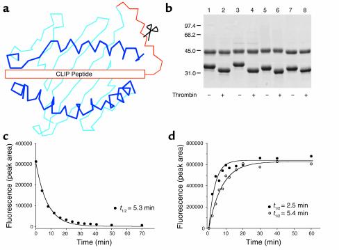
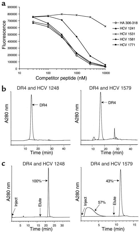
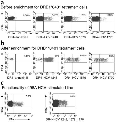
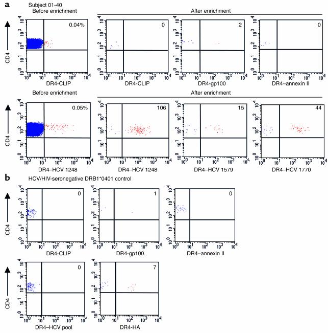
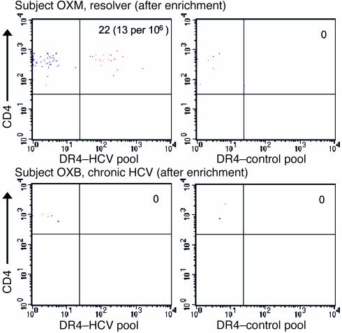
Containment of hepatitis C virus (HCV) and other chronic human viral infections is associated with persistence of virus-specific CD4 T cells, but ex vivo characterization of circulating CD4 T cells has not been achieved. To further define the phenotype and function of these cells, we developed a novel approach for the generation of tetrameric forms of MHC class II/peptide complexes that is based on the cellular peptide-exchange mechanism. HLA-DR molecules were expressed as precursors with a covalently linked CLIP peptide, which could be efficiently exchanged with viral peptides following linker cleavage. In subjects who spontaneously resolved HCV viremia, but not in those with chronic progressive infection, HCV tetramer-labeled cells could be isolated by magnetic bead capture despite very low frequencies (1:1,200 to 1:111,000) among circulating CD4 T cells. These T cells expressed a set of surface receptors (CCR7+CD45RA-CD27+) indicative of a surveillance function for secondary lymphoid structures and had undergone significant in vivo selection since they utilized a restricted Vbeta repertoire. These studies demonstrate a relationship between clinical outcome and the presence of circulating CD4 T cells directed against this virus. Moreover, they show that rare populations of memory CD4 T cells can be studied ex vivo in human diseases.


Figures

Figure 1
Figure 1
Generation of tetramers from MHC class II/CLIP precursors. (a) Recombinant HLA-DR molecules were expressed in which the CLIP peptide was covalently attached to the N-terminus of the DRβ chain. These precursors were converted to the peptide-receptive form by thrombin cleavage of the linker. This strategy allowed a series of tetramers to be generated from a precursor protein. (b) SDS-PAGE (10%) of purified DR/CLIP precursors (7 μg per lane). Four different DR molecules were expressed in which DRα was paired with one of four DRβ chains (lanes 1 and 2: DRB1*0101; lanes 3 and 4: DRB5*0101; lanes 5 and 6: DRB1*1501; lanes 7 and 8: DRB1*0401). Thrombin cleavage of the linker (lanes 2, 4, 6, and 8) removed the CLIP peptide and reduced the molecular weight of the DRβ chain. (c) Kinetics of CLIP dissociation from DR4 (DRA, DRB1*0401). DR4 molecules were incubated with an Alexa-488–labeled CLIP peptide, and dissociation of labeled CLIP was examined at different time points following addition of a molar excess of unlabeled HA (residues 306–318) peptide by separating DR-bound and free peptide on an HPLC gel filtration column. (d) Peptide association is rapid and follows the kinetics of CLIP dissociation. DR4 molecules were incubated for different time intervals with Alexa-488–labeled HA peptide. The kinetics of CLIP dissociation (t1/2 of 5.3 min) closely mirrored those of HA peptide association (t1/2 of 5.4 min) indicating rapid binding of peptide. Addition of DM further accelerated this process (t1/2 of 2.5 min).
Figure 2
Figure 2
Purification of HLA-DR molecules loaded with peptides from HCV. (a) Binding of HCV peptides to DR4. Based on a genome-wide analysis of the CD4 T cell response to HCV, seven candidate peptides were evaluated for DR4 binding. Unlabeled HCV peptides were used as unlabeled competitors for a biotinylated influenza HA (residues 306–318) peptide, and the amount of DR-bound biotinylated peptide was measured with europium-labeled streptavidin following capture of DR molecules with the L243 antibody. Two HCV peptides (HCV 1241 and HCV 1771; numbering based on the position of the first residue in the HCV polyprotein) showed a dose-response curve similar to that of the high-affinity influenza HA peptide, while higher concentrations of the HCV 1581 peptide were required for competition with labeled HA peptide. HCV 1531 is shown as an example of a peptide that did not effectively compete for binding of HA peptide to DR4. (b and c) Two-step purification of DR molecules loaded with defined peptides. DR4 molecules were separated from free peptide by HPLC gel filtration (b) and then injected into an HPLC DNP affinity column (c) to isolate DR4 molecules loaded with DNP-labeled HCV peptide. Bound complexes were eluted using 50 mM CAPS, pH 11.5, and immediately neutralized. A280 nm, absorbance measurements at a wavelength of 280 nm.
Figure 3
Figure 3
Staining of short-term HCV-specific CD4 T cell lines with MHC class II tetramers. (a) A short-term T cell line was generated from PBMCs by a single round of stimulation with 1 μg/ml of recombinant C200 antigen encoding the HCV NS3 and NS4 proteins. CD4 T cells were stained with PE-labeled MHC class II tetramers, including a control tetramer, DR4–annexin II, as well as three HCV tetramers, DR4–HCV 1248, DR4–HCV 1579, and DR4–HCV 1770. Cells were analyzed by flow cytometry prior to enrichment for PE-tetramer–positive cells; the percentage of CD4+/tetramer+ cells is indicated in the upper right quadrant. (b) Magnetic enrichment of tetramer-positive cells by positive selection of anti-PE–labeled cells. Data are shown for stimulated CD4 T cells from subject 01-40; similar data were obtained with short-term stimulated lines from subjects 99D and 98A. (c) Cells from an HCV NS3/NS4–stimulated line from subject 98A were stained with a pool of three HCV tetramers, DR4–HCV 1248, DR4–HCV 1579, and DR4–HCV 1770. Data are shown gated on CD4 T cells, without enrichment for tetramer-positive cells. Intracellular cytokine staining was performed by stimulation of cells from this same line with a pool of HCV peptides corresponding to the tetramer epitopes. Data are shown gated on CD4 T cells, and the percentage of IFN-γ+CD4+ cells is indicated in the upper right quadrant.
Figure 4
Figure 4
Ex vivo MHC class II tetramer staining of HCV-specific CD4 T cells. (a) PBMC from subject 01-40 with spontaneously resolved HCV viremia were stained with three control tetramers and three HCV tetramers. Cells were positively selected with anti-PE microbeads and analyzed by flow cytometry; the number of CD4+/tetramer+ cells is indicated in the upper right quadrant. An example of FACS analysis before enrichment for tetramer+ cells is shown for control tetramer DR4-CLIP and HCV tetramer DR4–HCV 1248. The frequencies of tetramer-positive cells in the CD4 T cell pool were determined by splitting the sample 9:1 following labeling with anti-PE beads; 90% of the cells were magnetically enriched, while 10% of the cells were analyzed without enrichment to determine the number of CD4 T cells. The frequencies of the tetramer+ cells of total CD4 cells are as follows: DR4–HCV 1248: 8.2 per 100,000 (0.008%); DR4–HCV 1579: 1.5 per 100,000 (0.0015%); DR4–HCV 1770: 2.4 per 100,000 (0.0024%). Representative data are shown from two independent experiments with fresh PBMC from subject 01-40. (b) PBMC from a DRB1*0401 HCV-seronegative control were stained with the following MHC class II tetramers: 3 control tetramers (DR4-CLIP, DR4-gp100, and DR4–annexin II), a pool of three HCV tetramers, and an HA (residues 306–318) tetramer. The number of CD4+/tetramer+ cells following enrichment with anti-PE microbeads is indicated in the upper right quadrant. The frequency of DR4–influenza HA–specific T cells in this subject was 0.9 per 100,000 (0.0009%).
Figure 5
Figure 5
Comparison of the T cell response to HCV in subjects with resolved viremia and chronic persistent infection. PBMCs from subject OXM (a spontaneous resolver) and OXB (chronic viremia) were labeled with a pool of the three HCV tetramers (left panels) or three control tetramers (DR4-CLIP, DR4-gp100, and DR4–annexin II). The number of CD4+/tetramer+ cells following enrichment with anti-PE microbeads is indicated in the upper right quadrant. HCV tetramer–positive CD4 T cells could not be detected in the four patients with chronic viremia (Table 2).
Figure 6
Figure 6
Ex vivo phenotypic analysis of HCV-specific memory CD4 T cells using MHC class II tetramers. (a) PBMCs from three subjects with resolved HCV viremia were stained with a pool of three DR4-HCV tetramers as well as antibodies to CCR7, CD45RA, or CD27. Tetramer-positive cells were positively selected with anti-PE microbeads and analyzed by flow cytometry. Plots are gated on CD4+/tetramer+ cells. (b) PBMCs from subject 01-40 were labeled with an antibody to CD62L in conjunction with the DR4–HCV 1579 and DR4–HCV 1770 tetramers.
Figure 7
Figure 7
TCR Vβ repertoire of HCV class II tetramer–positive cells. (a) Fresh PBMCs from subject 01-40 were stained with DR4–HCV 1248 tetramer, positively selected by labeling with anti-PE microbeads, and then split into separate tubes for staining with a panel of 16 Vβ antibodies. (b and c) Similarly, short-term stimulated HCV-specific CD4 T cell lines from subjects 01-40 (b) and 98A (c) were stained with DR4–HCV 1248 and subsequently stained with the 16 Vβ antibodies. The Vβ antibodies that positively stained DR4–HCV 1248+ cells are shown for each subject, as well as an example of a negative control Vβ antibody that did not stain the tetramer-positive cells. In subject 98A, the frequency of T cells in bulk PBMCs expressing Vβ5.1 and Vβ12 was 6.6% and 2.2% of CD4 T cells, respectively.

References

    1. Altman JD, et al. Phenotypic analysis of antigen-specific T lymphocytes. Science. 1996;274:94–96. - PubMed
    1. Meyer AL, et al. Direct enumeration of Borrelia-reactive CD4 T cells ex vivo by using MHC class II tetramers. Proc. Natl. Acad. Sci. U. S. A. 2000;97:11433–11438. - PMC - PubMed
    1. Homann D, Teyton L, Oldstone MB. Differential regulation of antiviral T-cell immunity results in stable CD8+ but declining CD4+ T-cell memory. Nat. Med. 2001;7:913–919. - PubMed
    1. Lauer GM, Walker BD. Hepatitis C virus infection. N. Engl. J. Med. 2001;345:41–52. - PubMed
    1. Alter MJ, et al. The prevalence of hepatitis C virus infection in the United States, 1988 through 1994. N. Engl. J. Med. 1999;341:556–562. - PubMed

Publication types