. 2003 Sep;112(6):843-52.
doi: 10.1172/JCI17647.

JunD protects against chronic kidney disease by regulating paracrine mitogens

Affiliations

JunD protects against chronic kidney disease by regulating paracrine mitogens

Evangéline Pillebout et al. J Clin Invest. 2003 Sep.

Abstract

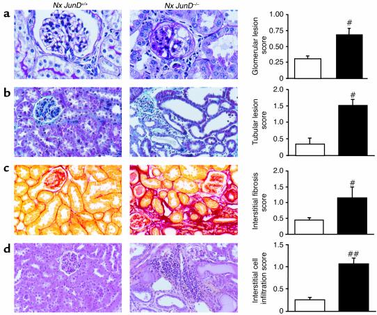
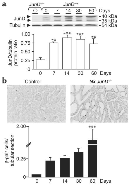
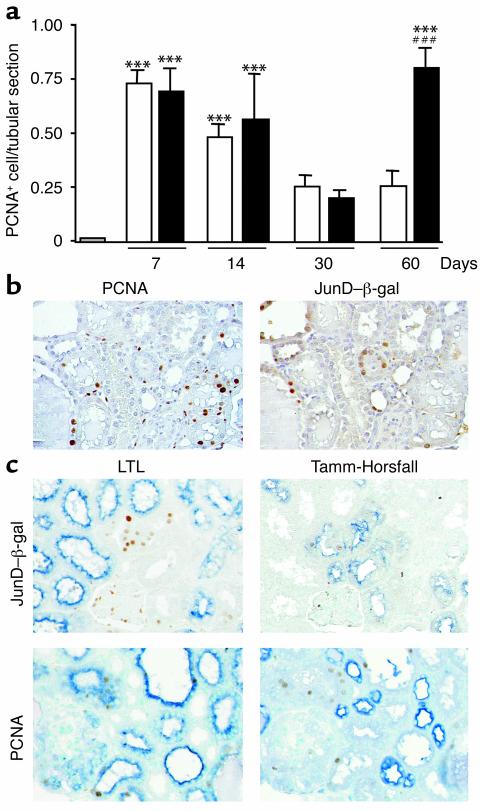
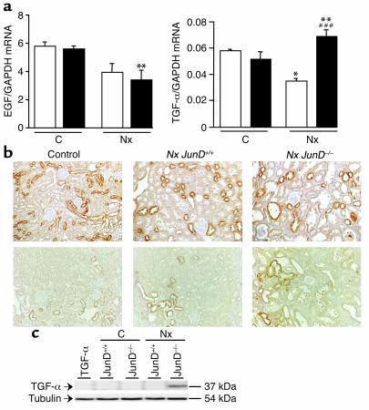
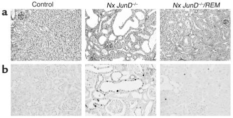
The AP-1 transcription factor, composed of Jun and Fos proteins, plays a crucial role in the fine tuning of cell proliferation. We showed previously that AP-1 complexes are activated during the proliferative response that parallels the development of renal lesions after nephron reduction, but little is known about the specific role of individual Jun/Fos components in the deterioration process. Here we used JunD knockout (JunD-/-) mice and an experimental model of chronic renal injury (75% nephron reduction) to explore the role of JunD. Nephron reduction resulted in an initial compensatory growth phase that did not require JunD. JunD, however, was essential to inhibit a second wave of cell proliferation and to halt the development of severe glomerular sclerosis, tubular dilation, and interstitial fibrosis. We show that the effects of junD inactivation are not cell autonomous and involve upregulation of the paracrine mitogen, TGF-alpha. Expression of a transgene (REM) encoding a dominant negative isoform of the EGFR, the receptor for TGF-alpha, prevented the second wave of cell proliferation and the development of renal lesions in bitransgenic JunD-/-/REM mice. We propose that JunD is part of a regulatory network that controls proliferation to prevent pathological progression in chronic renal diseases.


Figures

Figure 1
Figure 1
Accelerated renal lesion development in the absence of JunD. Morphology and lesion scores of remnant kidneys from JunD+/+ (white bars) and JunD–/– (black bars) mice 60 days after nephron reduction (Nx). Renal lesions were graded according to established methodology in randomly selected microscopic fields of the cortex. (a) Glomerular lesions, ×400. (b) Tubular lesions, ×200. (c) Interstitial fibrosis, ×200. (d) Interstitial mononuclear cell infiltration, ×200. Results represent mean ± SEM; n = 6. #P < 0.05, ##P < 0.01, JunD–/– versus JunD+/+ by Mann-Whitney test.
Figure 2
Figure 2
JunD expression increases after nephron reduction. (a) Immunoblot analysis of JunD expression in control nonoperated (designated 0) and remnant kidneys of JunD+/+ mice. Protein expression was analyzed before surgery (day 0) and 7, 14, 30, and 60 days after nephron reduction. Extracts of JunD–/– kidneys served as negative controls (C–). (b) Immunohistochemical analysis of JunD–β-gal reporter gene expression in control nonoperated (designated 0) and remnant kidneys of JunD–/– mice. β-gal staining was performed before surgery (day 0) and 7, 14, 30, and 60 days after nephron reduction. The top panels show staining in control and remnant kidneys at day 60 (× 200). The bottom panel shows the number of β-gal–positive cells per tubular section at each experimental time point. Data are mean ± SEM; n = 4–6 for each point. ANOVA, followed by Tukey-Kramer test: **P < 0.005, ***P < 0.001, remnant versus control kidneys.
Figure 3
Figure 3
Increased tubular cell proliferation in the absence of JunD. (a) The number of PCNA-positive cells per tubular section was measured in control kidneys (gray bar) and in JunD+/+ (white bars) and JunD–/– (black bars) remnant kidneys 7, 14, 30, and 60 days after nephron reduction. Data are mean ± SEM; n = 4–6 for each time point. ***P < 0.001, remnant versus control kidneys; ###P < 0.001, JunD–/– versus JunD+/+ by ANOVA, followed by Tukey-Kramer test. (b) PCNA and β-gal immunostaining in serial 4-μm sections of JunD–/– remnant kidneys at day 60 (×200). (c) Colocalization experiments in serial sections of JunD–/– remnant kidneys at day 60. Upper panels: overlay of β-gal staining and LTL staining (left) or Tamm-Horsfall staining (right). Lower panels: overlay of PCNA staining and LTL staining (left) or Tamm-Horsfall staining (right). (×200).
Figure 4
Figure 4
Elevated TGF-α expression in the absence of JunD. (a) EGF and TGF-α mRNA expression in control nonoperated (C) and remnant (Nx) kidneys of JunD+/+ (white bars) and JunD–/– (black bars) mice at day 60. Data are mean ± SEM; n = 6 for each point. *P < 0.05, **P < 0.01, remnant versus control kidneys; ###P < 0.001, JunD–/– versus JunD+/+ kidneys by ANOVA, followed by Tukey-Kramer test. (b) Immunohistochemistry of EGF (upper) and TGF-α (lower) expression in control nonoperated and remnant (Nx) kidneys of JunD+/+ and JunD–/– mice at day 60 (×200). (c) Immunoblot analysis of TGF-α expression in control nonoperated (C) and remnant (Nx) kidneys of JunD+/+ and JunD–/– mice at day 60. Extracts of TGF-α–/– kidneys served as negative controls.
Figure 5
Figure 5
Overexpression of a dominant negative EGFR prevents lesion development and cell proliferation in JunD–/– mice. (a) Morphology of control and of remnant (Nx) kidneys from JunD–/– and JunD–/–/REM mice at day 60. PAS staining, ×200. (b) Immunohistochemical analysis of PCNA in control and in remnant (Nx) kidneys from JunD–/– and JunD–/–/REM mice at day 60 (×200).

Similar articles

Cited by

References

    1. Hostetter TH. Progression of renal disease and renal hypertrophy. Annu. Rev. Physiol. 1995;57:263–278. - PubMed
    1. Olivetti G, Anversa P, Rigamonti W, Vitali-Mazza L, Loud AV. Morphometry of the renal corpuscle during normal postnatal growth and compensatory hypertrophy. A light microscope study. J. Cell Biol. 1977;75:573–585. - PMC - PubMed
    1. Terzi F, et al. Subtotal but not unilateral nephrectomy induces hyperplasia and protooncogene expression. Am. J. Physiol. 1995;268:F793–F801. - PubMed
    1. Kliem V, et al. Mechanisms involved in the pathogenesis of tubulointerstitial fibrosis in 5/6-nephrectomized rats. Kidney Int. 1996;49:666–678. - PubMed
    1. Terzi F, et al. Sodium restriction decreases AP-1 activation after nephron reduction in the rat: role in the progression of renal lesions. Exp. Nephrol. 2000;8:104–114. - PubMed

Publication types

MeSH terms