. 2003 Sep;112(6):883-91.
doi: 10.1172/JCI15483.

Renal protection from ischemia mediated by A2A adenosine receptors on bone marrow-derived cells

Affiliations

Renal protection from ischemia mediated by A2A adenosine receptors on bone marrow-derived cells

Yuan-Ji Day et al. J Clin Invest. 2003 Sep.

Abstract

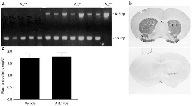
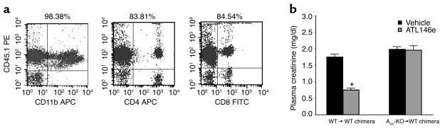
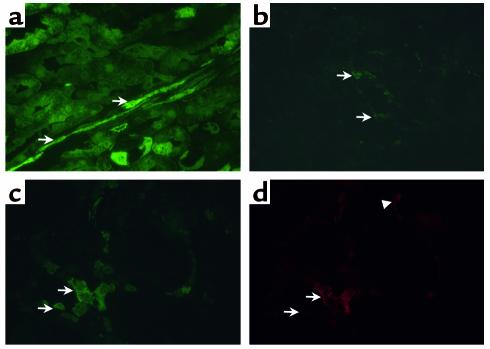
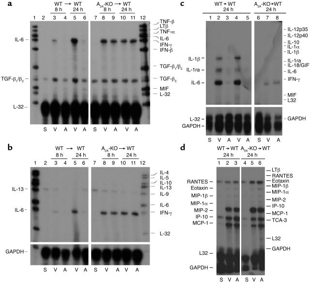
Activation of A2A adenosine receptors (A2ARs) protects kidneys from ischemia-reperfusion injury (IRI). A2ARs are expressed on bone marrow-derived (BM-derived) cells and renal smooth muscle, epithelial, and endothelial cells. To measure the contribution of A2ARs on BM-derived cells in suppressing renal IRI, we examined the effects of a selective agonist of A2ARs, ATL146e, in chimeric mice in which BM was ablated by lethal radiation and reconstituted with donor BM cells derived from GFP, A2AR-KO, or WT mice to produce GFP-->WT, A2A-KO-->WT, or WT-->WT mouse chimera. We found little or no repopulation of renal vascular endothelial cells by donor BM with or without renal IRI. ATL146e had no effect on IRI in A2A-KO mice or A2A-KO-->WT chimera, but reduced the rise in plasma creatinine from IRI by 75% in WT mice and by 60% in WT-->WT chimera. ATL146e reduced the induction of IL-6, IL-1beta, IL-1ra, and TGF-alpha mRNA in WT-->WT mice but not in A2A-KO-->WT mice. Plasma creatinine was significantly greater in A2A-KO than in WT mice after IRI, suggesting some renal protection by endogenous adenosine. We conclude that protection from renal IRI by A2AR agonists or endogenous adenosine requires activation of receptors expressed on BM-derived cells.


Figures

Figure 1
Figure 1
Genotyping and phenotyping mice for the A2AR gene. (a) Ethidium bromide–stained PCR amplification products of tail-clip DNA from WT (A2A+/+) and A2A-KO (A2A–/–) mice. The A2A-KO allele was identified by PCR of the inserted neomycin resistance cassette using oligonucleotide primers (see Methods) NEO-F and NEO-R and yielding a 618-bp product. The A2A WT allele was identified by PCR of a portion of the WT A2AR gene that was deleted from the KO construct using primers WT-F and WT-R and yielding a 163-bp band. +/+, WT; +/–, heterozygous; –/–, homozygous A2A-KO. Each lane represents PCR products from individual mice. (b) Immunohistochemical localization of A2AR in mouse forebrain sections. In the WT brain sections (top), dense A2AR-like immunoreactivity is present in the striatum (caudate putamen) and extends through the cell bridges of the striatum (arrow) to the olfactory tubercles on the ventral surface of the brain. In A2A-KO brain (bottom), specific immunoreactivity is completely absent. Scale bar, 1 mm. CPu, caudate putamen; Tu, tubercles; cc, corpus callosum. (c) Effect of ATL146e in A2A-KO mice. A2A-KO mice were subjected to IRI (see Methods). The rise in plasma creatinine after ATL146e was similar to that after vehicle administration (n = 6, not significant). Values are means ± SE.
Figure 2
Figure 2
Effect of renal IRI and ATL146e on plasma creatinine in chimeric mice. (a) Reconstitution efficacy of granulocytes and lymphocytes at 6 weeks after BM transplantation. PBMCs from transplanted recipients were purified and incubated with anti-CD45.1 as a marker of donor cells and either anti-CD11b, anti-CD4, or anti-CD8a monoclonal antibodies (see Methods). Labeled cells were enumerated by flow cytometry. The number at the top of each panel indicates the percentage of cells that are positive for both CD45.1 and each of the other marker fluorescent antibodies. Data are representative of 12 independent experiments. (b) Effect of renal IRI and ATL146e on plasma creatinine in chimeric mice. Chimeric mice were prepared by transferring to lethally irradiated WT recipients BM from either WT (WT→WT) or A2A-KO (A2A-KO→WT) donor animals. Chimeric animals were treated with vehicle or ATL146e (10 ng/kg per minute) beginning 5 hours before 32 minutes of ischemia and continuing for 24 hours of reperfusion. N = 11 for each group. Values are means ± SE. *P < 0.0001 versus vehicle.
Figure 3
Figure 3
Immunohistochemistry of GFP-positive cells in kidney of GFP mice and GFP→WT chimera. (a) Immunohistochemical localization of GFP in outer medulla of kidney from GFP mice. Magnification, ×630. Endothelial cells are identified by arrows. (b) Outer medulla of kidney from GFP→WT chimeras after renal IRI. Donor BM-derived cells are identified by arrows. Magnification, ×630. (c and d) Dual labeling of kidney from GFP→WT chimeras after renal IRI. Labeling with anti-GFP antibody (c) reveals cells of donor origin (arrows, c) that are identified as macrophages (arrows, d) by labeling with anti-macrophage antibody (d). Arrowhead indicates recipient macrophage. Magnification, ×1000 in (c) through (d).
Figure 4
Figure 4
Cytokine and chemokine gene expression from kidneys of chimeric mice subjected to IRI. Kidneys were subjected to 32 minutes of ischemia and either 8 or 24 hours of reperfusion in animals that were treated with either vehicle or ATL146e. V, vehicle; A, ATL146e. (a) RNase protection assay (RPA) analysis of selected cytokines from kidney mRNA using mCK3 probes. Sham-operated chimeric mice (lanes 2 and 7) and chimeric mice subjected to IRI (lanes 3–6 and 8–11). WT→WT mice at 8 (lanes 3 and 4) or 24 (lanes 5 and 6) hours and A2A-KO→WT mice at 8 (lanes 8 and 9) or 24 (lanes 10 and 11) hours as indicated. Each lane represents solution hybridization with RNA derived from kidneys of separate mice. (b) RPA analysis using the mCK1 probes. (c) RPA analysis using mCK2b probes. WT→WT mice at 24 hours (lanes 1–5) and A2A-KO→WT mice at 24 hours (lanes 6–8). Mice were subjected to sham operation (S) (lanes 1 and 6) or IRI (lanes 2–5, 7, and 8). Relative mobility of unprotected cytokine mRNA is illustrated on the right. S, sham operation. (d) RPA analysis using mCK5 probes. Kidney RNA was isolated from WT→WT mice at 24 hours (lanes 1–3) and from A2A-KO→WT mice at 24 hours (lanes 4–6). Mice were subjected to sham operation (lanes 1 and 4) or IRI (lanes 2, 3, 5, and 6).

Similar articles

Cited by

References

    1. Lauriat S, Linas SL. The role of neutrophils in acute renal failure. Semin. Nephrol. 1998;18:498–504. - PubMed
    1. Thadhani R, Pascual M, Bonventre JV. Acute renal failure. N. Engl. J. Med. 1996;334:1448–1460. - PubMed
    1. Zwacka RM, et al. CD4+ T-lymphocytes mediate ischemia/reperfusion-induced inflammatory responses in mouse liver. J. Clin. Invest. 1997;100:279–289. - PMC - PubMed
    1. Burne MJ, et al. Identification of the CD4+ T cell as a major pathogenic factor in ischemic acute renal failure. J. Clin. Invest. 2001;108:1283–1290. doi:10.1172/JCI200112080. - PMC - PubMed
    1. Rabb H, et al. Pathophysiological role of T lymphocytes in renal ischemia-reperfusion injury. Am. J. Physiol. Renal Physiol. 2000;279:F525–F531. - PubMed

Publication types

MeSH terms