. 2003 Sep;112(6):902-14.
doi: 10.1172/JCI18337.

Susceptible MHC alleles, not background genes, select an autoimmune T cell reactivity

Affiliations

Susceptible MHC alleles, not background genes, select an autoimmune T cell reactivity

Thomas Stratmann et al. J Clin Invest. 2003 Sep.

Abstract

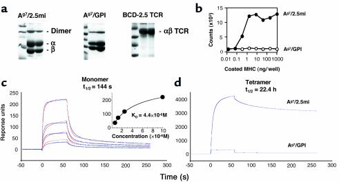
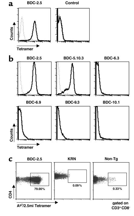
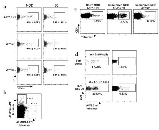
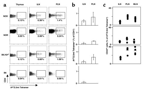
To detect and characterize autoreactive T cells in diabetes-prone NOD mice, we have developed a multimeric MHC reagent with high affinity for the BDC-2.5 T cell receptor, which is reactive against a pancreatic autoantigen. A distinct population of T cells is detected in NOD mice that recognizes the same MHC/peptide target. These T cells are positively selected in the thymus at a surprisingly high frequency and exported to the periphery. They are activated specifically in the pancreatic LNs, demonstrating an autoimmune specificity that recapitulates that of the BDC-2.5 cell. These phenomena are also observed in mouse lines that share with NOD the H-2g7 MHC haplotype but carry diabetes-resistance background genes. Thus, a susceptible haplotype at the MHC seems to be the only element required for the selection and emergence of autoreactive T cells, without requiring other diabetogenic loci from the NOD genome.


Figures

Figure 1
Figure 1
Biophysical and functional characterization of Ag7/2.5mi and Ag7/GPI MHC molecules. (a) SDS-PAGE analysis of the various recombinant MHC and TCR molecules used in this study. Molecules were purified from culture supernatants of transfected Drosophila melanogaster cells. The peak fractions of the final size exclusion chromatography are shown. (b) Recombinant Ag7/2.5mi molecules can activate the BDC-2.5 T cell hybridoma. Ag7/2.5mi MHC monomers were coated at the indicated concentrations into 96-well plates. IL-2 production was measured from the supernatants after 24 hours of culture. (c) Surface plasmon resonance analysis of the Ag7/2.5mi MHC/BDC-2.5 TCR interaction. Left: BDC-2.5 TCR molecules were randomly immobilized on a CM5 chip, and Ag7/2.5mi or Ag7/GPI (negative control) MHC molecules were flown over the surface. Subtracted curves are shown in blue lines, calculated curves in red. A Langmuir 1:1 binding model was used for analysis. Concentrations of injected MHC molecules were 10 μM, 2.5 μM, 1.25 μM, and 0.625 μM. The inset shows a steady-state analysis of the same interaction. (d) Tetramers of Ag7 with either peptide were flown over a high-density BDC-2.5 TCR surface. Unsubtracted curves corresponding to a tetramer concentration of 1 μM are presented.
Figure 2
Figure 2
Specific recognition of 2.5mi+ T cells by Ag7/2.5mi multimers. (a) BDC-2.5 T cell hybridoma and 19.2B T cell hybridoma (specific for glutamic acid decarboxylase peptide 282–292) were labeled with either Ag7/2.5mi tetramers (solid line) or Ag7/GPI tetramers (dotted line). (b) Staining of T cell clones specific for β cell granule membranes with Ag7/2.5mi tetramers. After 4 days of stimulation in vitro with islet-membrane preparations, BDC clones were stained with Ag7/2.5mi tetramers. (c) Inguinal LN cells from young adult BDC-2.5/NOD, KRN, and NOD (Non-Tg) mice were stained for CD4 and Ag7/2.5mi tetramers.
Figure 3
Figure 3
Presence of 2.5mi+ T cells in NOD mice. (a) Specific 2.5mi+CD4+ T cells were identified in subcutaneous LNs from young adult NOD but not from B6 mice. Control stains for either Ag7/GPI or I-Ag7/HEL tetramers are shown in the lower panels. (b) Lack of cross-reactivity of Ag7/2.5mi tetramers with nonspecific T cells. Splenocytes from a NOD mouse were stained with PE-labeled Ag7/2.5mi tetramers, followed by incubation with APC-labeled Ag7/GPI tetramers. T cells were only stained by either of the tetramers, not by both simultaneously, indicating specific recognition. (c) 2.5mi+CD4+ T cells expansion after immunization with the 2.5 mi+ peptide. Popliteal LN cells from nonimmunized and immunized NOD mice were stained with either Ag7/2.5mi or Ag7/GPI tetramers. (d) Expansion of 2.5mi+CD4+ cells in a lymphopenic mouse reconstituted with 2.5mi+CD4+ cells (or 2.5miCD4+ cells as control), sorted from splenocytes of 5-week-old NOD mice and immediately analyzed for sort purity. Twenty thousand sorted cells were injected into RAGo/o/NOD mice. Inguinal LN cells were stained 20 days after transfer for the presence of 2.5mi+CD4+ cells. The number of injected cells was not corrected for the presence of nonviable cells and is therefore likely to be overestimated. The number of 2.5mi+CD4+ cells present in the whole mouse (n) was extrapolated to a full lymphoid compartment from the number of 2.5mi+CD4+ cells observed in the inguinal LN by flow cytometry (taking into account multiple losses during the staining procedures and the flow cytometry acquisition itself).
Figure 4
Figure 4
Histological detection of 2.5mi+CD4+ T cells. In situ staining of 2.5mi+CD4+ T cells in PLNs (a and b) and pancreatic islets (ce). I-Ag7/2.5mi MHC tetramer stains are shown in green (a and c). Staining with an anti-CD4 antibody is shown in red and overlaid by the tetramer staining (b and d). Insulin staining is shown in e.
Figure 5
Figure 5
T cells with the 2.5mi+ reactivity are positively selected in the thymus and expand in the periphery. (a and b) Thymocytes, splenocytes, inguinal LNs (ILNs), PLNs, and infiltrated pancreatic T cells were stained for CD4, CD8, CD3, and Ag7/2.5mi tetramer. The percentage of Ag7/2.5mi+CD4+CD3hi T cells was electronically calculated from the rectangle gate of each profile. (c) 2.5mi+CD4+ T cells expand in the periphery after leaving the thymus, and they accumulate in the PLNs. Average values represent independent experiments for seven mice analyzed. (d) Specific activation of 2.5mi+CD4+ T cells in PLNs. ILNs, PLNs, or superior mesenteric LNs (MLNs) from BDC-2.5 transgenic (Tg) mice (open squares) or from groups of 3-week-old (diamonds) or 4-week-old or older NOD mice (triangles) were costained with Ag7/2.5mi tetramers and anti-CD44.
Figure 6
Figure 6
Time course analysis of 2.5mi+ T cells in naive NOD females. PLNs and iliac LNs (ILLNs) were removed and analyzed for 2.5mi+ T cells at the indicated ages. Harvested lymphocytes were pooled from four animals before analysis.
Figure 7
Figure 7
Reactivity of 2.5mi+ T cell hybridomas to 2.5mi peptide and pancreatic islet cells. T cell hybridoma clones obtained from NOD females after immunization with 2.5mi peptide (a and c) or from naive animals (b and c) were incubated in the presence of APCs with increasing concentrations of 2.5mi peptide (a and b) or pancreatic islet cells (c), and their IL-2 response was measured by incorporation of 3H-thymidine during an NK cell proliferation assay. Clone numbers were as follows: (a) A42, open squares; A162.7, filled circles; A202.2, filled squares; A64, open diamonds; A129, filled triangles; A129, filled diamonds; A72, open upward-pointing triangles; A86, open circles; and A166, open downward-pointing triangles; (b) C26, open squares; C20, open circles; C33, filled upward-pointing triangles; C87, filled circles; BDC-2.5, filled squares; C36, open upward-pointing triangles; C1, filled downward-pointing triangles; C26, open downward-pointing triangles.
Figure 8
Figure 8
Ag7 is sufficient to select 2.5mi+CD4+ T cells. (a) Thymocytes, inguinal LNs (ILNs), and PLNs from young adult NOD mice, NOR mice, C57BL/6 mice congenic for H-2g7 (B6.H2g7), and C57BL/6 (B6) mice were stained for CD4, CD8, CD3, and Ag7/2.5mi tetramer. The percentage of Ag7/2.5mi+CD4+CD3hi T cells was electronically calculated from the rectangle gate of each profile. (b) 2.5mi+ T cells accumulate in PLNs of mice that do not get insulitis or diabetes but share Ag7. Average representation (percent) of 2.5mi+CD4+ T cells from NOD, NOR, and B6.H2g7 mice (five to seven mice per group) was measured in four independent experiments. (c) 2.5mi+ T cells are activated in PLNs of mice that do not get insulitis or diabetes but share Ag7. The percentage of CD44hiCD4+Ag7/2.5mi+ T cells in ILNs, PLNs, and MLNs of NOD, NOR, and B6.H2g7 mice (five to seven mice per group) was measured in four independent experiments.

Comment in

Similar articles

Cited by

References

    1. Tisch R, McDevitt H. Insulin-dependent diabetes mellitus. Cell. 1996;85:291–297. - PubMed
    1. Vyse TJ, Todd JA. Genetic analysis of autoimmune diabetes. Cell. 1996;85:311–318. - PubMed
    1. Wicker LS, Todd JA, Peterson L. Genetic control of autoimmune diabetes in the NOD mouse. Annu. Rev. Immunol. 1995;13:179–200. - PubMed
    1. Hattori M, et al. The NOD mouse: recessive diabetogenic gene in the major histocompatibility complex. Science. 1986;231:733–735. - PubMed
    1. Acha-Orbea H, McDevitt HO. The first external domain of the nonobese diabetic mouse class II I-A beta chain is unique. Proc. Natl. Acad. Sci. U. S. A. 1987;84:2435–2439. - PMC - PubMed

Publication types

MeSH terms