. 2003 Oct;1(1):E13.
doi: 10.1371/journal.pbio.0000013. Epub 2003 Sep 15.

Drosophila free-running rhythms require intercellular communication

Affiliations

Drosophila free-running rhythms require intercellular communication

Ying Peng et al. PLoS Biol. 2003 Oct.

Abstract

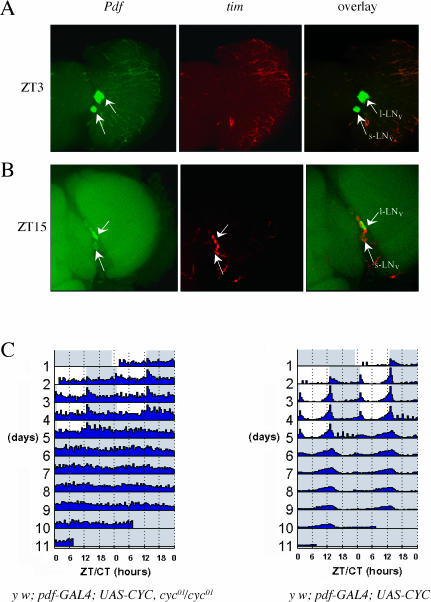
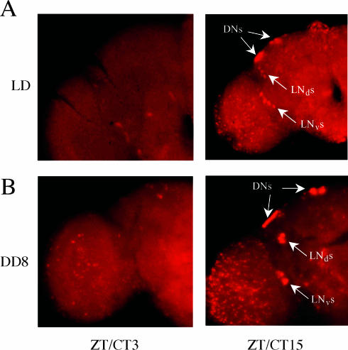
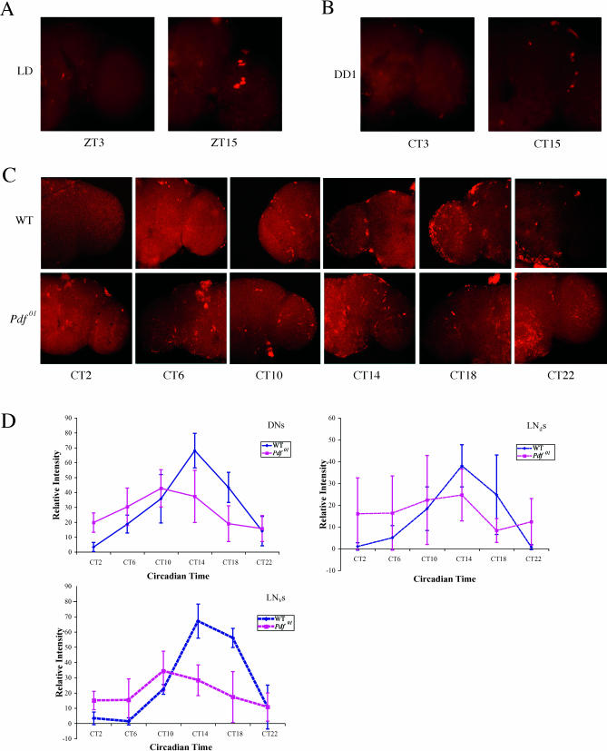
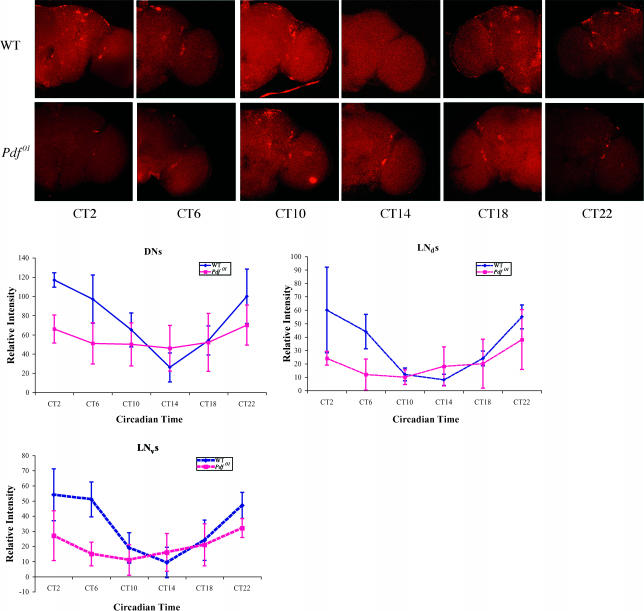
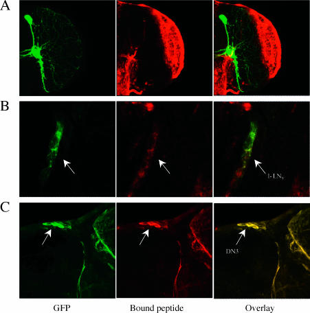
Robust self-sustained oscillations are a ubiquitous characteristic of circadian rhythms. These include Drosophila locomotor activity rhythms, which persist for weeks in constant darkness (DD). Yet the molecular oscillations that underlie circadian rhythms damp rapidly in many Drosophila tissues. Although much progress has been made in understanding the biochemical and cellular basis of circadian rhythms, the mechanisms that underlie the differences between damped and self-sustaining oscillations remain largely unknown. A small cluster of neurons in adult Drosophila brain, the ventral lateral neurons (LN(v)s), is essential for self-sustained behavioral rhythms and has been proposed to be the primary pacemaker for locomotor activity rhythms. With an LN(v)-specific driver, we restricted functional clocks to these neurons and showed that they are not sufficient to drive circadian locomotor activity rhythms. Also contrary to expectation, we found that all brain clock neurons manifest robust circadian oscillations of timeless and cryptochrome RNA for many days in DD. This persistent molecular rhythm requires pigment-dispersing factor (PDF), an LN(v)-specific neuropeptide, because the molecular oscillations are gradually lost when Pdf(01) mutant flies are exposed to free-running conditions. This observation precisely parallels the previously reported effect on behavioral rhythms of the Pdf(01) mutant. PDF is likely to affect some clock neurons directly, since the peptide appears to bind to the surface of many clock neurons, including the LN(v)s themselves. We showed that the brain circadian clock in Drosophila is clearly distinguishable from the eyes and other rapidly damping peripheral tissues, as it sustains robust molecular oscillations in DD. At the same time, different clock neurons are likely to work cooperatively within the brain, because the LN(v)s alone are insufficient to support the circadian program. Based on the damping results with Pdf(01) mutant flies, we propose that LN(v)s, and specifically the PDF neuropeptide that it synthesizes, are important in coordinating a circadian cellular network within the brain. The cooperative function of this network appears to be necessary for maintaining robust molecular oscillations in DD and is the basis of sustained circadian locomotor activity rhythms.


Conflict of interest statement

The authors have declared that no conflicts of interest exist.

Figures

Figure 1
Figure 1. Rescuing Molecular Oscillations within the LNvs Is Not Sufficient to Rescue Locomotor Activity Rhythms
The rescued mutant genotype is y w;pdf–GAL4;UAS–CYC,cyc01/cyc01. The flies were entrained in standard LD conditions and timepoints taken. Molecular oscillations were examined by whole-mount in situ hybridization of the tim gene. Double staining with a Pdf probe was used to label the LNvs neuronal group. (A and B) These show representative duplicate experiments. No tim mRNA signal is detectable in the dorsal region of the brain. The lower arrows point to the s-LNvs and the upper arrows to the l-LNvs. (A) Brain taken at timepoint ZT3. Panels shown from left to right are Pdf (green, FITC labeled), tim (red, Cy3 labeled), and an image overlay. (B) Brain taken at timepoint ZT15. Panels shown from left to right are Pdf (green, FITC labeled), tim (red, Cy3 labeled), and an image overlay. (C) The double-plotted actograms of rescue mutant and control flies in a standard LD:DD behavior assay. The colors on the background indicate the lighting conditions of the behavior monitors (white, lights on; light blue, lights off). In the actogram, the average locomotor activity of the group of flies is plotted as a function of time. The left panel shows the actogram of the rescued mutant flies (y w;pdf–GAL4/+;UAS–CYC,cyc01/cyc01, n = 30). RI (rhythm index; Levine et al. 2002a) = 0.14. The right panel shows the actogram for the rescued wild-type (control) flies (y w;pdf–GAL4/+;UAS–CYC/+, n = 32, RI = 0.61).
Figure 2
Figure 2. All Brain Clock Neuronal Groups Maintain Robust Oscillations of tim RNA Levels in DD
Wild-type flies were entrained for at least 3 days and then released into DD. tim RNA was assayed at trough (left panels) and peak (right panels) timepoints by whole-mount in situ hybridization. Wild-type flies in LD (A) were compared with the eighth day of DD (B). On the eighth day of DD, the locomotor activities of the fly population were still in close synchrony, without any obvious phase spreading (data not shown). Left panels, brains at ZT3 (A) or CT3 (B); right panels, brains from ZT15 (A) or CT15 (B). Both (A) and (B) are representative of three replicate experiments.
Figure 3
Figure 3. Molecular Oscillations of tim RNA Damp in DD in the Pdf01 Mutant
tim RNA oscillations were examined in the Pdf01 mutant under both LD (A) and different days in DD ([B] and [C]), by whole-mount in situ hybridization. (A), (B), and (C) are representative images from replicas of three experiments. (A) The left panel is from ZT3, and the right panel is from ZT15. A normal tim oscillation profile is observed compared to that of wild-type (see Figure 2A). (B) Brains from the Pdf01 mutant in the first day of DD. Left panel, CT3; right panel, CT15. Oscillations are comparable to those in LD. (C) Brains taken in the fourth day of DD. Six timepoints were taken throughout the circadian day. The sequence of panels from left to right is CT2, 6, 10, 14, 18, and 20, respectively. Wild-type brains (top row) were assayed in parallel with those from the Pdf01 mutant (bottom row). See text for details. (D) Quantification of (C). Relative intensities are taken from normalized mean pixel intensities. Different clock neuronal groups were quantified independently and compared between wild-type (blue curves) and Pdf01 mutant (purple curves). The panels from left to right are quantification of tim RNA oscillation in the DNs, in the LNds, and in the LNvs. Reduced cycling amplitude and a significant advanced phase were observed in the fourth day of DD. See text for details.
Figure 4
Figure 4. cry RNA Oscillation Amplitude Is Also Reduced by the Fourth Day of DD in the Pdf01 Mutant
cry RNA expression in the brain was examined at the fourth day of DD by whole-mount in situ hybridization using a cry probe. Timepoints were taken every 4 hours throughout the circadian day. The sequence of panels from left to right is CT2, 6, 10, 14, 18, and 20, respectively. Wild-type brains (top row) were analyzed in parallel with those from the Pdf01 mutant (bottom row). Shown are representative images from duplicate experiments. Quantification of cry RNA oscillations in different cell groups is as shown in Figure 3. Ubiquitous damping of the cycling amplitude in the different cell groups was observed in the Pdf01 mutant.
Figure 5
Figure 5. A PDF Peptide Binds to Many Cells, Including Several Clock Neuronal Groups
In vitro biontinylated PDF peptide was used to visualize the peptide binding locations (middle panels, with Cy3) in the brain (see Materials and Methods for details). We used membrane-bound GFP (green panels on the left) to label specific circadian neurons as well as their projections (right panels show the overlay of both channels). (A) The brain is from flies with labeled LNvs (y w,UAS–mCD8iGFP;pdf–GAL4). Numerous cells at the periphery of the medulla have the vast majority of the bound PDF peptide signal within the brain. This region receives widespread dendritic arborizations from the l-LNvs. (B) Bound PDF peptide was also detected on the surface of LNvs at a lower intensity. LNv cell bodies were labeled using UAS–mCD8iGFP;pdf–GAL4. Since the signal from the Cy3 channel was much weaker than the GFP signal, we reduced the output gain from the GFP channel. Sequential scanning was used to prevent cross-talk between the two channels. (C) y w,UAS–mCD8iGFP;tim–GAL4/+ flies were used to label all circadian neurons. In the dorsal region shown in this series, the arrow points to a group of DN3 neurons.

Similar articles

Cited by

References

    1. Allada R, White N, So W, Hall J, Rosbash M. A mutant Drosophila homolog of mammalian Clock disrupts circadian rhythms and transcription of period and timeless . Cell. 1998;93:791–804. - PubMed
    1. Allada R, Emery P, Takahashi JS, Rosbash M. Stopping time: The genetics of fly and mouse circadian clocks. Ann Rev Neurosci. 2001;24:1091–1119. - PubMed
    1. Allada R, Kadener S, Nandakumar N, Rosbash M. A recessive mutant of Drosophila Clock reveals a role in circadian rhythm amplitude. EMBO J. 2003;22:3367–3375. - PMC - PubMed
    1. Bae K, Lee C, Hardin PE, Edery I. dCLOCK is present in limiting amounts and likely mediates daily interactions between the dCLOCK–CYC transcription factor and the PER–TIM complex. J Neurosci. 2000;20:1746–1753. - PMC - PubMed
    1. Balsalobre A, Damiola F, Schibler U. A serum shock induces circadian gene expression in mammalian tissue culture cells. Cell. 1998;93:929–937. - PubMed

Publication types

MeSH terms

LinkOut - more resources