

Protein synthesis by single ribosomes
- PMID: 13130131
- PMCID: PMC1370481
- DOI: 10.1261/rna.5800303
Protein synthesis by single ribosomes
Abstract
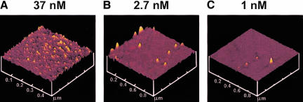
The ribosome is universally responsible for synthesizing proteins by translating the genetic code transcribed in mRNA into an amino acid sequence. Ribosomes use cellular accessory proteins, soluble transfer RNAs, and metabolic energy to accomplish the initiation, elongation, and termination of peptide synthesis. In translocating processively along the mRNA template during the elongation cycle, ribosomes act as supramolecular motors. Here we demonstrate that ribosomes adsorbed on a surface, as for mechanical or spectroscopic studies, are capable of polypeptide synthesis and that tethered particle analysis of fluorescent beads connected to ribosomes via polyuridylic acid can be used to estimate the rate of polyphenylalanine synthesis by individual ribosomes. This work opens the way for application of biophysical techniques, originally developed for the classical motor proteins, to the understanding of protein biosynthesis.
Figures


Similar articles
-
Rate of elongation of polyphenylalanine in vitro.Eur J Biochem. 1982 Feb;122(1):193-7. doi: 10.1111/j.1432-1033.1982.tb05866.x. Eur J Biochem. 1982. PMID: 7037399
-
Elongation factor G-promoted translocation and polypeptide elongation in ribosomes without GTP cleavage: use of columns with matrix-bound polyuridylic acid.Methods Enzymol. 1979;60:761-79. doi: 10.1016/s0076-6879(79)60070-8. Methods Enzymol. 1979. PMID: 379541 No abstract available.
-
Ribosomal synthesis of polyleucine on polyuridylic acid as a template: contribution of the elongation factors.FEBS Lett. 1980 Oct 20;120(1):135-40. doi: 10.1016/0014-5793(80)81064-7. FEBS Lett. 1980. PMID: 7002608 No abstract available.
-
Translation of genetic information on the ribosome.Angew Chem Int Ed Engl. 1971 Sep;10(9):638-51. doi: 10.1002/anie.197106381. Angew Chem Int Ed Engl. 1971. PMID: 5001418 Review. No abstract available.
-
Protein synthesis.Annu Rev Biochem. 1973;42:397-438. doi: 10.1146/annurev.bi.42.070173.002145. Annu Rev Biochem. 1973. PMID: 4581230 Review. No abstract available.
Cited by
-
Visualization of mRNA translation in living cells.J Cell Biol. 2006 Oct 9;175(1):67-76. doi: 10.1083/jcb.200512137. J Cell Biol. 2006. PMID: 17030983 Free PMC article.
-
Enzymatic Synthesis and Fractionation of Fluorescent PolyU RNAs.Bio Protoc. 2018 Sep 5;8(17):e2988. doi: 10.21769/BioProtoc.2988. eCollection 2018 Sep 5. Bio Protoc. 2018. PMID: 34395788 Free PMC article.
-
Multiple LacI-mediated loops revealed by Bayesian statistics and tethered particle motion.Nucleic Acids Res. 2014;42(16):10265-77. doi: 10.1093/nar/gku563. Epub 2014 Aug 12. Nucleic Acids Res. 2014. PMID: 25120267 Free PMC article.
-
Ribosomal protein L3 functions as a 'rocker switch' to aid in coordinating of large subunit-associated functions in eukaryotes and Archaea.Nucleic Acids Res. 2008 Nov;36(19):6175-86. doi: 10.1093/nar/gkn642. Epub 2008 Oct 2. Nucleic Acids Res. 2008. PMID: 18832371 Free PMC article.
-
DNA looping kinetics analyzed using diffusive hidden Markov model.Biophys J. 2007 Apr 15;92(8):L64-6. doi: 10.1529/biophysj.107.104828. Epub 2007 Feb 2. Biophys J. 2007. PMID: 17277177 Free PMC article.
References
-
- Alexander, R.W., Muralikrishna, P., and Cooperman, B.S. 1994. Ribosomal components neighboring the conserved 518–533 loop of 16S rRNA in 30S subunits. Biochemistry 33: 12109–12118. - PubMed
-
- Ban, N., Nissen, P., Hansen, J., Moore, P.B., and Steitz, T.A. 2000. The complete atomic structure of the large ribosomal subunit at 2.4 Å resolution. Science 289: 905–920. - PubMed
-
- Czworkowski, J. and Moore, P.B. 1997. The conformational properties of elongation factor G and the mechanism of translocation. Biochemistry 36: 10327–10334. - PubMed
-
- Frank, J. 2001. Cryo-electron microscopy as an investigative tool: The ribosome as an example. BioEssays 23: 725–732. - PubMed
-
- Howard, J. 2001. Mechanics of motor proteins and the cytoskeleton. Sinauer Associates, Inc., Sunderland, MA.
Publication types
MeSH terms
Substances
Grants and funding
LinkOut - more resources
Full Text Sources
Other Literature Sources