. 2003 Oct;9(10):1221-31.
doi: 10.1261/rna.5111803.

Nonreplicative homologous RNA recombination: promiscuous joining of RNA pieces?

Affiliations

Nonreplicative homologous RNA recombination: promiscuous joining of RNA pieces?

Anatoly P Gmyl et al. RNA. 2003 Oct.

Abstract

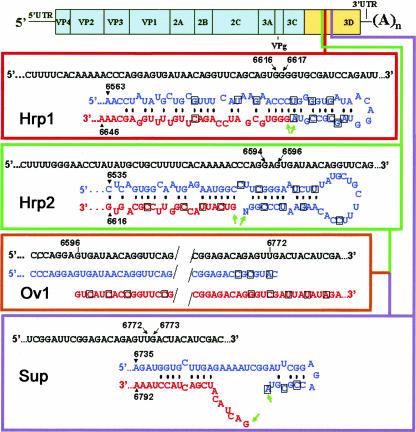
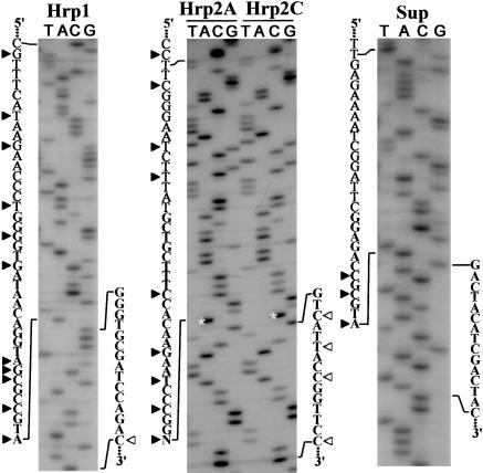
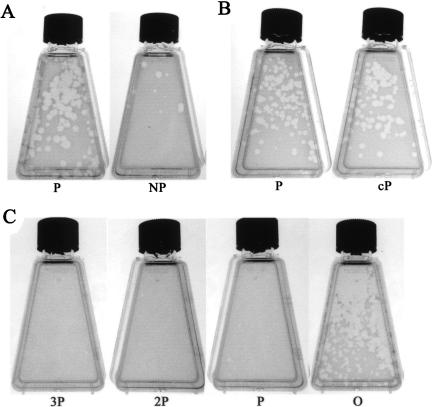
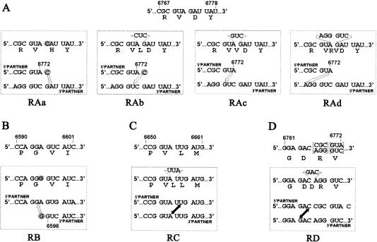
Biologically important joining of RNA pieces in cells, as exemplified by splicing and some classes of RNA editing, is posttranscriptional, whereas in RNA viruses it is generally believed to occur during viral RNA polymerase-dependent RNA synthesis. Here, we demonstrate the assembly of precise genome of an RNA virus (poliovirus) from its cotransfected fragments, which does not require specific RNA sequences, takes place before generation of the viral RNA polymerase, and occurs in different ways: Apparently unrestricted ligation of the terminal nucleotides, joining of any one of the two entire fragments with the relevant internal nucleotide of its partner, or internal crossovers within the overlapping sequence. Incorporation of the entire 5' or 3' partners into the recombinant RNA is activated by the presence of terminal 3'-phosphate and 5'-OH, respectively. Such postreplicative reactions, fundamentally differing from the known site-specific and structurally demanding cellular RNA rearrangements, might contribute to the origin and evolution of RNA viruses and could generate new RNA species during all stages of biological evolution.


Figures

FIGURE 1.
FIGURE 1.
Structure of the recombination partners. The viral RNA is composed of the 5UTR, polyprotein reading frame, 3UTR, and poly(A). The sequence encoding viral RNA polymerase (3D) is in yellow. The wild-type nucleotide sequences corresponding to the junction between the terminal sequences of the recombination partners are shown as upper lines in each rectangle, with the coordinates of the partners’ termini indicated. Terminal sequences of the 5′- and 3′-segments are in blue and red, respectively, with engineered silent mutations boxed. The 3′-terminal N of Hrp2 corresponds to the nucleotide, A or C, ligated to the original transcript. The 5′- and 3′-Ov1 partners correspond to the 5′ Sup and 3′ Hrp2 partners (with additional silent mutations), respectively. The green arrows mark the to-be-ligated nucleotides. Possible heteroduplex structures between the partners in Hrp1 and Hrp2 pairs are shown. There are no stable heteroduplexes in the case of Sup or Ov1 pairs (although some potential pairing is shown for the former case).
FIGURE 2.
FIGURE 2.
Ligation of fragments of the viral RNA. Examples of sequences at the crossover sites of the ligation products of different pairs of RNA fragments. For the Hrp2 pair, the results with the variants 3′-terminated with A (Hrp2A) and C (Hrp2C) are shown. The 5′- and 3′-terminal sequences of the respective ligation partners are given at the left and right sides of each sequencing gel, respectively. The silent mutations serving as genetic markers of the 5′- and 3′-partners are shown as solid and empty arrowheads, respectively. The bands corresponding to different 3′-terminal nucleotides of the Hrp2A and Hrp2C 5′-fragments are marked with asterisks.
FIGURE 3.
FIGURE 3.
Plaque formation by the ligation products of the genomic segments with different terminal structures. (A) Ligation of the P and NP forms of the Sup 5′-partner with the O form of its counterpart. (B) Ligation of the P and cP forms of the Hrp2C 5′-partner with the O form of its counterpart. (C) Ligation of the P form of the Sup 5′-partner with the O, P, 2P, and 3P forms of its counterpart. The plaques were photographed on the third day after transfection.
FIGURE 4.
FIGURE 4.
Three types of genomes generated by recombination between Ov1 partners. Terminal sequences of the partners are shown on the left side of the panel. The 3′-terminal C-residue of the 5′-partner, which was removed during preparation of its P form, is shown in parentheses. The genetic markers of the 5′- and 3′-partners are shown as solid and empty arrowheads, respectively. Portions of the sequencing gels corresponding to the termini of the partners are shown. Arrows indicate the direction of electrophoresis. Examples of recombinants obtained upon crossing NP and 3P (internal crossover), NP and O (entire 3′-partner), and P and 3P (entire 5′-partner) forms are given.
FIGURE 5.
FIGURE 5.
Aberrant recombination between Ov1 partners involving incorporation of the entire 5′-partner (A), or 3′-partner (B), or internal crossovers (C,D). The expected (and actually most often found) nucleotide and amino acid sequences of the crossover regions of the relevant precise recombinants are shown above the rectangles. Amino acids are given in the single-letter code. Because the parts of the overlapping regions in the 5′- and 3′-partners shown in panel D are different due to the presence of silent marker mutations, the bracketed codons in the precise recombinant might be represented by two synonymous versions. The determined aberrant sequences are given in the upper parts of the rectangles. Nucleotides corresponding to the point mutations in recombinants RAa and RB are shadowed; insertions of three or six nucleotides in all other recombinants are indicated. Proposed mechanisms underlying generation of the aberrant recombinants are given in the lower parts of the rectangles. The additional terminal nucleotides presumably added by T7 polymerase to the 5′-partner (RAa and RAb) and 3′-partner (RB) are encircled. Open arrows connect a terminal nucleotide of the 5′-partner or 3′-partner with an internal nucleotide of their counterparts; double-headed solid arrows connect the ligated nucleotides in internal crossovers.
FIGURE 6.
FIGURE 6.
“Monstrous” recombinant genomes. (A) Schematic structures of the recombinant RNAs obtained in separate experiments involving the Hrp1 pair, in which a partially degraded 3′-partner was used. Portions originating from the 5′- and 3′-partners are shown in black and gray, respectively, with coordinates of the segments derived from the 3′-partner indicated. The stars mark the correct fusion between positions 6616 and 6617. The arrowheads denote the terminal codon of the polyprotein frame. Segments corresponding to the untranslated regions are shown thinner than the coding regions. (B) Instability of the SK132 genome. The PCR products obtained by using primers corresponding to positions 6496–6515 (sense) and 5′ (T)15CTCC 3′ (antisense) of the Hrp1 RNA from the newly generated SK132 genome and from the viral RNA harvested from its four consecutive passages in Vero cells. Sequencing of the 958-nt-long and 1066-nt-long products revealed the structures corresponding to the original Hrp1 and the monster shown in panel A, respectively.

Similar articles

Cited by

References

    1. Abelson, J., Trotta, C.R., and Li, H. 1998. tRNA splicing. J. Biol. Chem. 273: 12685–12688. - PubMed
    1. Agol, V.I. 1997. Recombination and other genomic rearrangements in picornaviruses. Semin. Virol. 8: 1–9.
    1. Agol, V.I. 2002. Picornavirus genetics: An overview. In The molecular biology of picornaviruses (eds. B.L. Semler and E. Wimmer), pp. 269–284. ASM Press, Washington, DC.
    1. Arnold, J.J. and Cameron, C.E. 1999. Poliovirus RNA-dependent RNA polymerase (3Dpol) is sufficient for template switching in vitro. J. Biol. Chem. 274: 2706–2716. - PubMed
    1. Avota, E., Berzins, V., Grens, E., Vishnevsky, Y., Luce, R., and Biebricher, C. 1998. The natural 6 S RNA found in Qβ-infected cells is derived from host and phage RNA. J. Mol. Biol. 276: 7–17. - PubMed

Publication types