. 1992 Sep;59(3):1145-52.
doi: 10.1111/j.1471-4159.1992.tb08357.x.

Evidence for the implication of phosphoinositol signal transduction in mu-opioid inhibition of DNA synthesis

Affiliations

Evidence for the implication of phosphoinositol signal transduction in mu-opioid inhibition of DNA synthesis

J Barg et al. J Neurochem. 1992 Sep.

Abstract

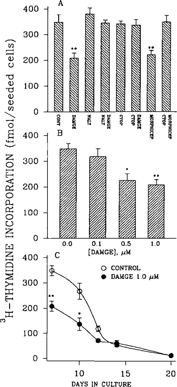
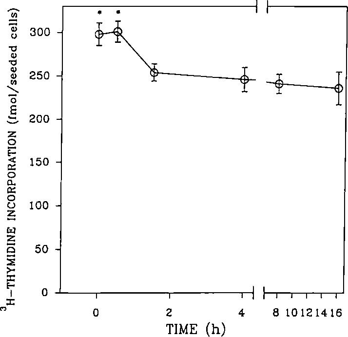
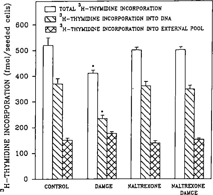
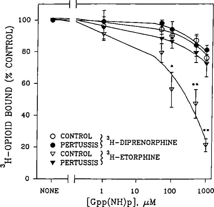
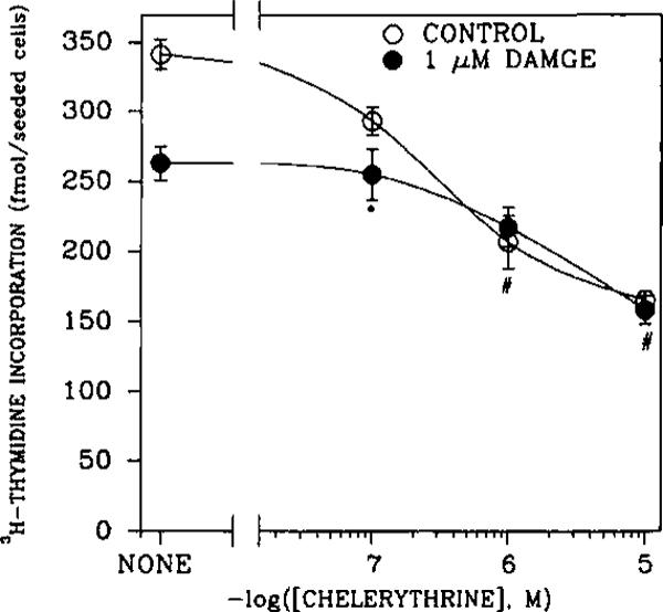
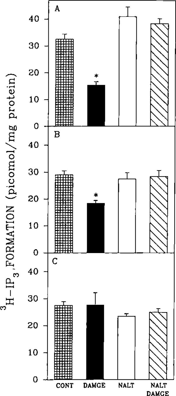
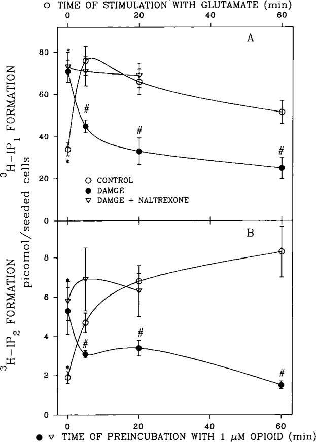
An opioid receptor agonist, [D-Ala2,Me-Phe4,Glyol5]enkephalin (DAMGE), decreased [3H]thymidine incorporation into DNA of fetal rat brain cell aggregates. This action proved to depend on the dose of this enkephalin analog and the interval the aggregates were maintained in culture. The opioid antagonist naltrexone and the mu-specific antagonist cyclic D-Phe-Cys-Tyr-D-Trp-Orn-Thr-Pen-Thr amide (CTOP) reversed the DAMGE effect, arguing for a receptor-mediated mechanism. The mu-opioid nature of this receptor was further established by inhibiting DNA synthesis with the highly mu-selective agonist morphiceptin and blocking its action with CTOP. Several other opioids, pertussis toxin, and LiCl also diminished DNA synthesis, whereas cholera toxin elicited a modest increase. Naltrexone completely reversed the inhibition elicited by the combination of DAMGE and low doses of LiCl but not by that of high levels of LiCl alone. The enkephalin analog also reduced basal [3H]inositol trisphosphate and glutamate-stimulated [3H]inositol monophosphate and [3H]inositol bisphosphate accumulation in the aggregates. These DAMGE effects were reversed by naltrexone and were temporally correlated with the inhibition of DNA synthesis. A selective protein kinase C inhibitor, chelerythrine, also inhibited thymidine incorporation dose-dependently. The effect of DAMGE was not additive in the presence of chelerythrine but appeared to be consistent with their actions being mediated via a common signaling pathway. These results suggest the involvement of the phosphoinositol signal transduction system in the modulation of thymidine incorporation into DNA by DAMGE.


Figures

FIG. 1
FIG. 1
[3H]Thymidine incorporation into DNA of rat whole-brain cell aggregates. A: Effect of 1 μM DAMGE, morphiceptin (MORPHICEP), CTOP, and naltrexone (NALT) on [3H]thymidine (106 dpm per plate) incorporation into DNA of 7-day brain cell aggregates. CONT, control. B: Dose-dependent DAMGE inhibition of [3H]thymidine incorporation into DNA of 7-day brain cell aggregates. C: Effect of DAMGE on [3H]thymidine incorporation into DNA of rat whole-brain cell aggregates as a function of days in culture. Data are mean ± SE (bars) values from three to five experiments performed in duplicate. *p < 0.05, **p < 0.01, compared with the control.
FIG. 2
FIG. 2
Preincubation time dependence of DAMGE inhibition of [3H]thymidine incorporation into DNA of rat brain cell aggregates. Cultures were preincubated for the indicated periods with 1 μM DAMGE and washed three times with 40 ml of Eagle's medium. For the last 23 h [3H]thymidine was added, and incorporation was determined for the 7-day culture. Data are mean ± SE (bars) values from three experiments. *p < 0.05 for significance of difference from other time intervals.
FIG. 3
FIG. 3
[3H]Thymidine incorporation into DNA and non-DNA cell compartments of rat fetal brain cell aggregates. Samples were divided into two aliquots, and [3H]thymidine incorporation into each was determined as described in Materials and Methods. Total [3H]thymidine incorporation into cells was estimated after washing intact aggregates with phosphate-buffered saline (4 × 20 ml). The difference between total incorporation into the cell and the amount incorporated into DNA represents the intracellular thymidine and is referred to as the external pool. Data are mean ± SE (bars) values from three experiments. *p < 0.05 for significance of difference from other treatments.
FIG. 4
FIG. 4
Modulation of [3H]thymidine incorporation into DNA of rat brain cell aggregates. A Concentration-dependent effects of LiCl or pertussis toxin on DAMGE (1 μM) inhibition of [3H]thymidine (106 dpm per plate) incorporation into DNA of whole-brain cell aggregates. Cultures were exposed to opioids, toxins, or LiCl 48 h before harvesting and to [3H]thymidine for the final 23 h and analyzed after 7 days of culture. B: Dose-dependent naltrexone reversal of the inhibition of [3H]thymidine incorporation into DNA by 10 mM LiCl, 1 μM DAMGE in the presence of 0.5 mM LiCl, or 5 ng/ml of pertussis toxin in 7-day whole-brain cell aggregates. C: Dose-dependent cholera toxin potentiation of [3H]thymidine incorporation into DNA of control or DAMGE (1 μM)-treated 7-day whole-brain cell aggregates. Data are mean ± SE (bars) values from three to seven experiments. *p < 0.05, **p < 0.01 for significance of difference from the controls (cells untreated by the agent listed in the abscissa of each panel).
FIG. 5
FIG. 5
Concentration dependence of Gpp(NH)p inhibition of opioid agonist and partial agonist specific binding to opioid receptors in rat brain cell aggregates. Membranes from pertussis toxin-treated (5 ng/ml, 48 h) or control cells were incubated with 1 nM [3H]diprenorphine or [3H]etorphine in the presence or absence of indicated concentrations of Gpp(NH)p. Specific binding of cells untreated with Gpp(NH)p was > 1,300 dpm per tube. Data are mean ± SE (bars) values from three experiments. *p < 0.05, **p < 0.01 for significance of difference from the pertussis toxin-treated aggregates.
FIG. 6
FIG. 6
Effect of the PKC inhibitor chelerythrine and DAMGE (1 μM) on [3H]thymidine incorporation into DNA in 7-day brain cell aggregates. Aggregates were exposed to chelerythrine and/or opioid 48 h before harvesting and to [3H]thymidine for the final 23 h of 7-day cultures. Data are mean ± SEM (bars) values from three to six experiments. *p < 0.05 for significance of difference from cultures treated only with chelerythrine (control). #p < 0.05 for significance of difference for cultures not treated with chelerythrine.
FIG. 7
FIG. 7
Effect of opioid agonist and antagonist on [3H]IP3 accumulation in whole-brain cell aggregates. A: Effect of DAMGE and/or naltrexone (NALT; 1 μM) on [3H]IP3 accumulation in 7-day cultures. B: Effect of DAMGE and/or naltrexone (1 μM) on [3H]IP3 accumulation in 10-day cultures. C: Effect of DAMGE and/or naltrexone (1 μM) on [3H]IP3 accumulation in 14-day rat fetal brain cell aggregates. Data are mean ± SE (bars) values from three to seven experiments. *p < 0.05 for difference from control (CONT).
FIG. 8
FIG. 8
Effect of opioid agonist and antagonist on glutamate-promoted (A) [3H]inositol monophosphate ([3H]IP2) and (B) [3H]-inositol bisphosphate ([3H]IP2) accumulation in whole-brain cell aggregates. Aggregates (7 days) were preincubated with 1 μM DAMGE with or without 1 μM naltrexone for 0−60 min and then treated with 40 μM glutamate for 0−60 (A) or 20 (B) min. The cells were harvested, and 3H-IP accumulation was measured as described in Materials and Methods. Data are mean ± SE (bars) values from three to seven experiments. Zero-time values are significantly different from those of all other time points: *p < 0.05. Values for glutamate-stimulated IP accumulation in the presence of DAMGE are significantly different from those obtained in its absence: *p < 0.05.

Similar articles

Cited by

References

    1. Ashkenazi A, Ramachandran J, Capon DJ. Acetylcholine analogue stimulates DNA synthesis in brain-derived cells via specific muscarinic receptor subtypes. Nature. 1989;340:146–150. - PubMed
    1. Barg J, Levy R, Simantov RJ. Paradoxical and subtype-specific effects of opiate antagonists on the expression of opioid receptors in rat brain cultures. J. Neurosci. Res. 1989a;22:322–330. - PubMed
    1. Barg J, Levy R, Simantov R. Expression of the three opioid receptor subtypes μ, δ and κ in guinea pig and rat brain cell cultures and in vivo. Int. J. Dev. Neurosci. 1989b;7:173–180. - PubMed
    1. Barg J, Belcheva M, Bern WT, Lambourne B, McLachlan JA, Tolman KC, Johnson FE, Coscia CJ. Desipramine modulation of sigma and opioid peptide receptor expression in glial cells. Peptides. 1991;12:845–849. - PubMed
    1. Bartolome JV, Bartolome MB, Daltner LA, Evans CJ, Barchas JD, Kuhn CM, Schanberg SM. Effects of β-endorphin on ornithine decarboxylase in tissues of developing rats. A potential role for this endogenous neuropeptide in the modulation of tissue growth. Life Sci. 1986;38:2355–2362. - PubMed

Publication types

MeSH terms