

doi: 10.1161/01.cir.12.2.259.
Chronic heart block in dogs; a method for producing experimental heart failure
- PMID: 13240809
- PMCID: PMC2950642
- DOI: 10.1161/01.cir.12.2.259
Item in Clipboard
Chronic heart block in dogs; a method for producing experimental heart failure
Circulation.
1955 Aug.
Abstract
A method is described for the production of chronic atrioventricular block in dogs, by incision of the region of the bundle of His through the open right atrium during temporary caval occlusion. Exercise tolerances, chest x-ray films, electrocardiograms, phonocardiograms, cardiac outputs, intracardiac pressures, femoral pressures, and left ventricular coronary flows were obtained preoperatively and from 1 to 10 months postoperatively. The majority of the animals developed clinical, laboratory, and pathological evidence of congestive heart failure. All animals had generalized myocardial hypertrophy.
Figures
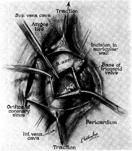
Artist's view of the technique for surgical production of complete heart block. The auricle has been entered during temporary caval occlusion, and the cut made across the auriculoventricular junction at the posterior end of the base of the septal cusp.

Details of the anatomy of the intracardiac incision for heart block. The auricles have been removed, and the view is from above. Note the relation of the cut to the posterior end of the septal cusp, the left auricle, and the base of the aorta.

Two-meter chest x-ray films preoperatively (left), and 12 days after performance of surgical complete heart block (right). Preoperative rate was 120 per minute and idioventricular rate was 40 per minute. Note cardiac enlargement and pulmonary vascular congestion after block.

Spectral phonocardiogram of dog which had been completely blocked for four months. The auricular sounds (a) seen in the record were easily detectable upon stethoscopic examination, although softer than the regular heart sounds (1, 2).
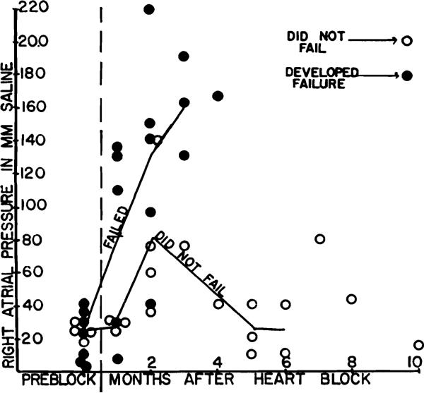
Right atrial pressures in dogs with complete heart block, the open circles representing measurements from animals which did not fall, and the black dots being from animals which decompensated.

Femoral arterial A, right ventricular B, and right atrial C pressures. Calibrations of the first two are in millimeters Hg and of the third in millimeters water. The records are all from animals in heart failure.

Mean femoral arterial pressures in animals with chronic complete heart block, expressed in per cent of preoperative controls (each animal thus serving as its own control). Note lack of correlation of values to presence or absence of heart failure.

Exercise tolerances in dogs with complete heart block, expressed in per cent of preoperative treadmill times. Points shown by solid circles are from animals which developed heart failure, and points shown by open circles are from animals which did not decompensate.

Cardiac output in animals with complete heart block expressed in per cent of preoperative values (each animal serving as his own control). All pre- and postoperative determinations were done with 27-30 mg. per kilogram. Nembutal except the eight week tests which were done with 20 to 23 mg. per kilogram. With the lighter anesthesia note the inability of the animals which were failing or later failed to increase their output as markedly as the compensated animals.
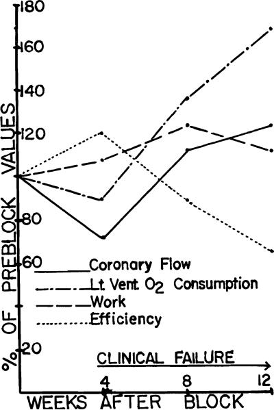
Left ventricular coronary flow, work, oxygen consumption, and efficiency before (four week determination), and after the onset of congestive heart failure (8 and 12 week determinations). Note the progressive fall in efficiency after decompensation, from a level which was initially higher than obtained in the preoperative control.

Total heart weights of dogs with complete heart block, in per cent of values predicted by method of Herrmann. Note absence of correlation between the degree of hypertrophy and the presence or absence of heart failure.

The ratio of left to right ventricular weight (L/R) in per cent of the values predicted by the method of Herrmann. The changes in left (Lt. Vent.) and right ventricular (Rt. Vent.) weight are also shown. Solid circles represent left ventricular weights, crosses represent L/R ratios, and open circles represent right ventricular weights.

Sectioris of the liver (upper) and lung (lower) showing central lobular atrophy and pulmonary edema. (liver (upper) × 75 lung (lower)–× 150).
References
-
- Goodale WT, Hackel DB. Measurement of coronary blood flow in dogs and man from rate of myocardial nitrous oxide desaturation. Circulation Research. 1953;1:502. - PubMed
-
- Kety S, Schmidt CF. The determination of cerebral blood flow in man by the use of nitrous oxide in low concentrations. Am. J. Physiol. 1945;143:53.
-
- Erlanger J, Blackman JR. A study of relative rhythmicity and conductivity in various regions of the auricles of the mammalian heart. Am. J. Physiol. 1907;19:125.
-
- Eyster JAE, Swarthout EC. Experimental determination of the influence of abnormal cardiac rhythms on the mechanical efficiency of the heart. Arch. Int. Med. 1920;25:317.
MeSH terms
Grants and funding
LinkOut - more resources
Full Text Sources
