. 1992 Mar;101 ( Pt 3)(Pt 3):701-7.
doi: 10.1242/jcs.101.3.701.

The 9E3 protein: immunolocalization in vivo and evidence for multiple forms in culture

Affiliations

The 9E3 protein: immunolocalization in vivo and evidence for multiple forms in culture

M Martins-Green et al. J Cell Sci. 1992 Mar.

Abstract

The avian gene 9E3/CEF4 belongs to a group of genes whose products are highly conserved and are homologous to inflammatory mediators. These genes, sometimes referred to as the gro family, are also expressed upon wounding or serum-stimulation of quiescent cells, suggesting that they may be important in aspects of growth and/or wound healing. We have used an antibody to the product of the 9E3 gene to show for the first time the distribution in vivo of the protein of one of these genes. The polyclonal antibody was produced against a synthetic peptide, [Cys76], 9E3, (77-103), located at the carboxy end of the molecule. The specificity of the antibody was determined by transfection of the 9E3 cDNA into Cos 7 cells, which do not express this gene. Moreover, despite the high homology between 9E3 and IL-8, the antibody did not crossreact with this molecule. The antibody was used to immuno-precipitate the protein from cultured normal and RSV-transformed chick embryo fibroblasts (CEFs) and to determine its distribution in tissues of newly hatched chicks. The staining was abundant in the cells and extracellular matrix (ECM) of connective tissue and other tissues of mesenchymal origin, such as bone and tendon. Most cells in the granulation tissue of wounds stained, some more intensely than others; the ECM also stained, especially in areas of scar tissue where collagen is abundant. In RSV-induced tumors, the protein was absent except in necrotic areas where a few cells--potentially macrophages--stained. In general, as expected, the protein was present in the cells and tissues that expressed the mRNA, but there were exceptions.(ABSTRACT TRUNCATED AT 250 WORDS)


Figures

Fig. 1
Fig. 1
Immunoblot analysis of the fractions collected after elution of the 9E3 antibody from the peptide-conjugated Affigel column. Most of the antibody was eluted in fractions 3 and 4. (W) The peptide was incubated with whole immune serum; (F) with the immune serum after incubation with the peptide-resin conjugate; and (C) without antibody.
Fig. 2
Fig. 2
Immunostaining of Cos 7 cells transfected with the 9E3 gene. (A, B) Cells transfected with the vector containing the gene in the sense direction and (C, D) controls consisting of cells with the gene in the antisense direction. (A) In situ hybridization with an antisense 9E3 mRNA-labeled probe showing the grains (arrowheads) over the cells. (B) Immunolabeling with the 9E3 antibody using alkaline phosphatase (arrowheads). (C) Same as A, showing no grains over the antisense-transfected cells. (D) Same as B, showing no staining of the antisense-transfected cells. Bar, 50 μm.
Fig. 3
Fig. 3
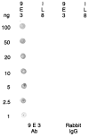
Immunoblot analysis of the cross reactivity of the 9E3 antibody with IL-8. The anti-9E3 antibody (350 ng/ml) did not recognize IL-8. The control consisted of rabbit IgG at the same concentration as the antibody for 9E3.
Fig. 4
Fig. 4
Immunostaining of cultured CEFs with the 9E3 antibody. (A) Starved confluent CEFs show staining in some of the cells and in the ECM associated with them. (B) RSV-transformed CEFs show intense staining especially in rounded cells. Bar, 20 μm.
Fig. 5
Fig. 5
Immunoprecipitation of the 9E3 protein from cultured cells. (A) Conditioned medium from confluent starved CEFs; (B) cell extracts from confluent starved CEFs; (C) conditioned medium from transformed cells and (D) cell extracts from transformed cells. (E–H) Controls: repeat of A–D but the immunoprecipitations were performed with nonimmune rabbit IgG at the same concentration as the 9E3 antibody. Numbers indicate the Mr (×10−3) of the marker proteins.
Fig. 6
Fig. 6
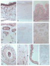
Immunostaining of normal wing tissues of newly hatched chicks with the 9E3 antibody. (A–C) Skin: (A) the fibroblasts of the dermis stain variably, with some labeling strongly (arrowheads). Staining is also visible in the ECM between the cells. The epidermis, especially the stratum corneum, stains intensely even though in situ hydridization with a radioactively labeled mRNA probe shows that it does not have mRNA at detectable levels (C). (D–F) Bone: (D) the osteoblasts in the lacunae and the osteocytes in less mature areas (arrowheads) stain intensely for 9E3, whereas the osteocytes in more mature areas do not stain (double arrowheads). (F) In situ hybridization with a radioactively labeled mRNA probe shows that the cells labeling with the antibody also label for the mRNA (grains). (G) Artery showing that the fibroblasts (arrowheads) of the connective tissue surrounding the smooth muscle layer (intima) stain very intensely; the endothelial cells do not stain (arrows) and the intima layer (stars) contains blotchy staining. (H) Skeletal muscle showing that the muscle fibers do not stain but cells of the connective tissue that surround them do (arrowheads). (I) Tendon showing that many fibroblasts (arrowheads) and their ECM stain intensely. (J) Cells of the bone marrow do not stain for 9E3. Controls (B, E) were done by incubating adjacent sections with the antibody preincubated with excess peptide for 3 h at RT. Bar, 50 μm.
Fig. 7
Fig. 7
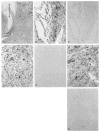
Immunostaining of wounded tissues of newly hatched chicks. (A) An overview of the granulation tissue at the wound site showing the general distribution of staining. Letters indicate areas enlarged in B, D, F and the two-headed arrow represents where the wounding clip was placed. (B, D, F) Virtually all cells stain for 9E3 except the endothelial cells (arrows). As in normal tissues, some cells stain more intensely than others. Note that in many cases the stain is also present in the ECM (arrowheads). (C, E, G) Controls performed as described for normal tissues. Bar, 50 μm.
Fig. 8
Fig. 8
Immunostaining of tumor tissues with the 9E3 antibody. (A) Healthy tumor tissue does not stain for 9E3. (B) Necrotic areas, however, contain cells (arrowheads) that stain for the 9E3 protein. (C) These necrotic areas also contain cells that stain for peroxidases (arrowheads), suggesting that they could be macrophages. (D) Control for the peroxidase reaction; the sections were incubated exactly as in C but in the absence of substrate. Bar, 50 μm.

Similar articles

Cited by

References

    1. Anlsowicz A, Bardwell L, Sager R. Constitutive overexpression of a growth-regulated gene in transformed Chinese hamster and human cells. Proc Nat Acad Sci USA. 1987;84:7188–7192. - PMC - PubMed
    1. Anlsowkz A, Zajchowskl D, Stenman G, Sager R. Functional diversity of gro gene expression in human fibroblasts and mammary epithelial cells. Proc Nat Acad Sci USA. 1988;85:9645–9649. - PMC - PubMed
    1. Appella E, Matushima K, Oppenhelm JJ, Yoshlmura T, Leonard EJ, Clore GM, Gronenborn AM. Determination of the primary and secondary structure of NAP-1/IL-8 and a monocyte chemoattractant protein, MCP-l/MCAF. Prog Clin Biol Res. 1990;349:405–417. - PubMed
    1. Baggiollnl M, Walz A, Kunkel SL. Neutrophil-activating Peptide-1/Interlukin 8, a novel cytokine that activates neutrophils. J Clin Invest. 1989;84:1045–49. - PMC - PubMed
    1. Baldwin ET, Weber IT, St Charles R, Xuan JC, Appella E, Yamada M, Matsushima K, Edwards BFP, Cloew GM, Gronenborn AM, Wlodawer A. Crystal Structure of interleukin 8: Symbiosis of NMR and crystallography. Proc Nat Acad Sci USA. 1991;88:502–506. - PMC - PubMed

Publication types