Comparative Study
. 1992;2(3):241-57.

Tissue-specific expression of the skeletal alpha-actin gene involves sequences that can function independently of MyoD and Id

Affiliations
Comparative Study

Tissue-specific expression of the skeletal alpha-actin gene involves sequences that can function independently of MyoD and Id

G E Muscat et al. Gene Expr. 1992.

Abstract

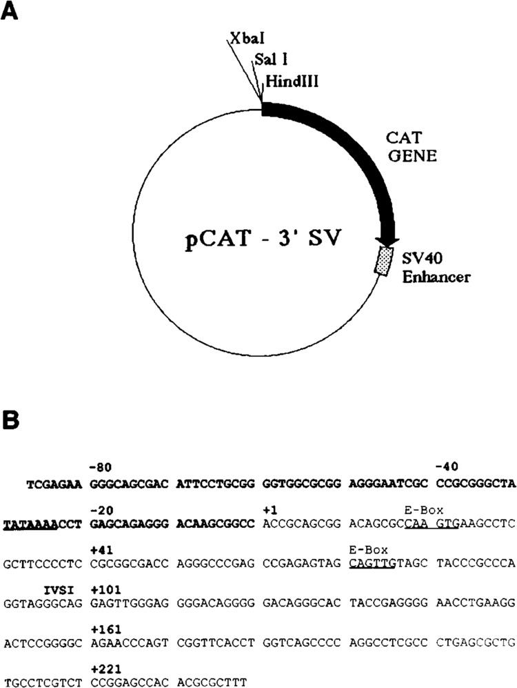
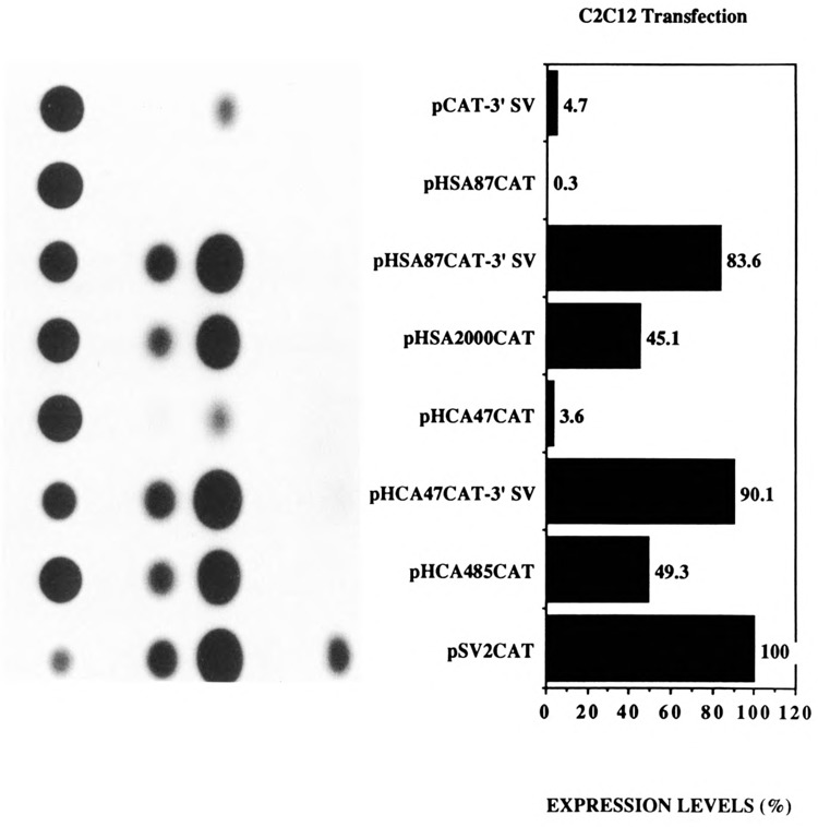
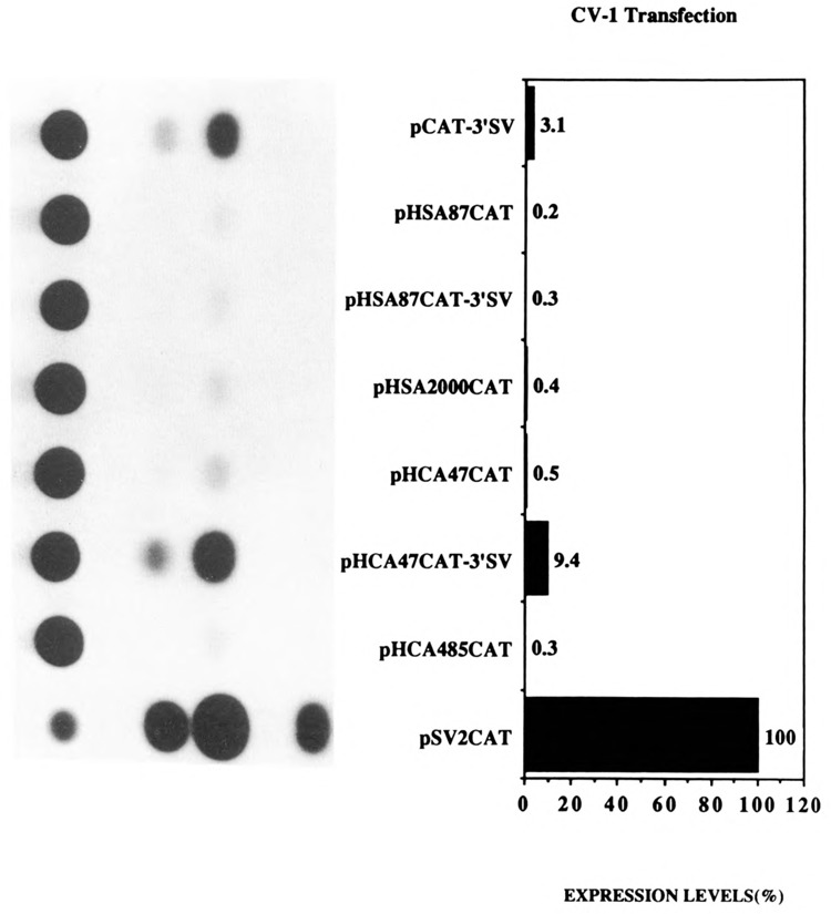
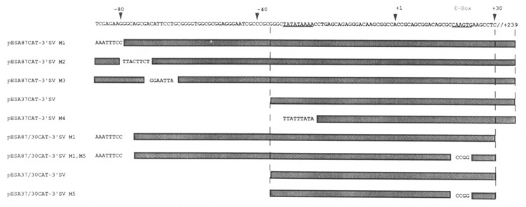
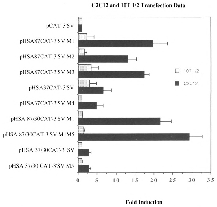
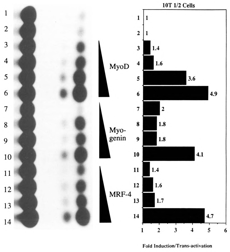
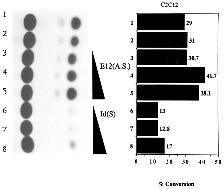
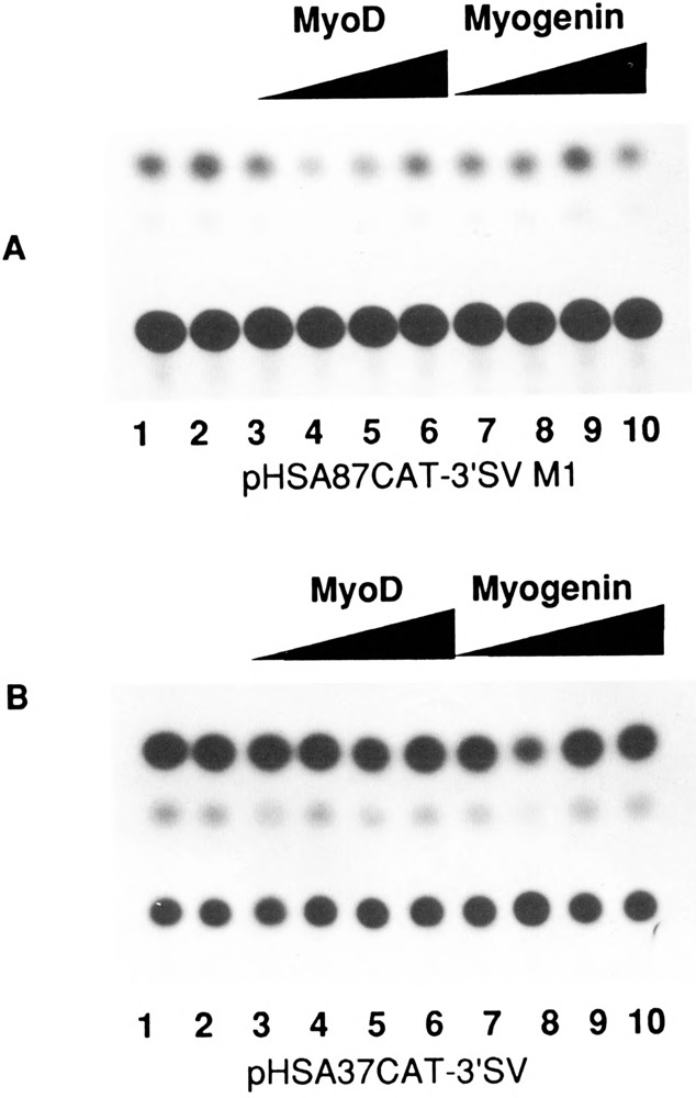
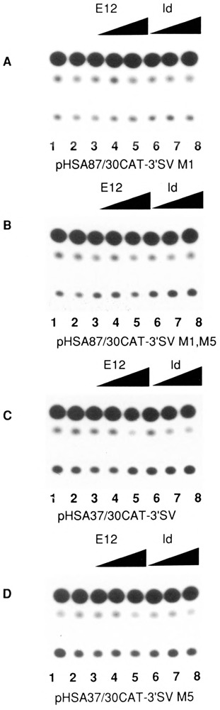
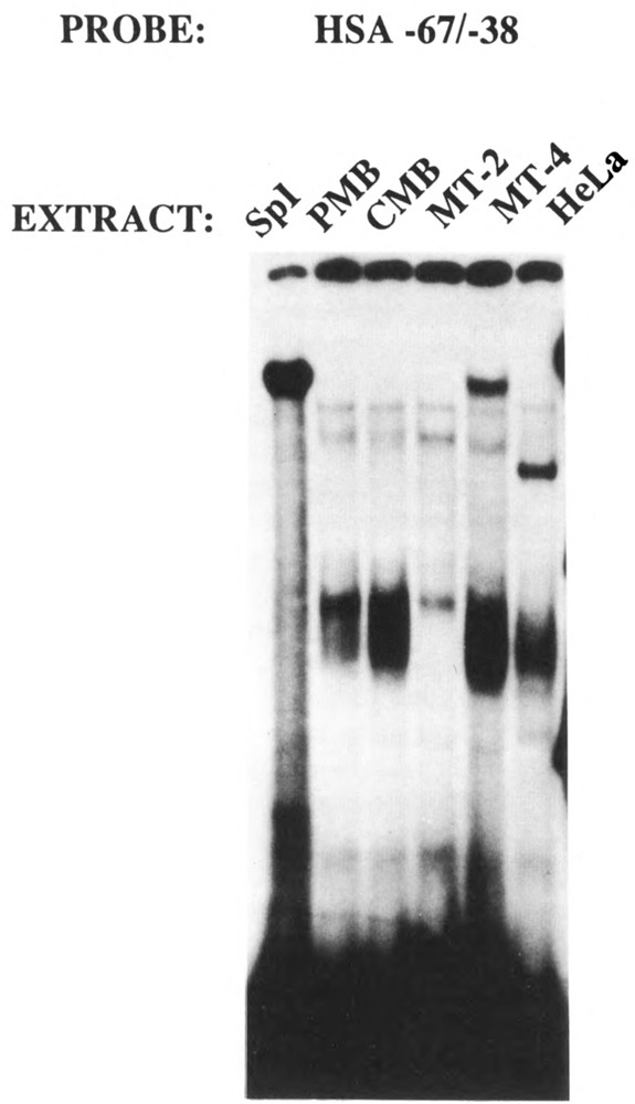
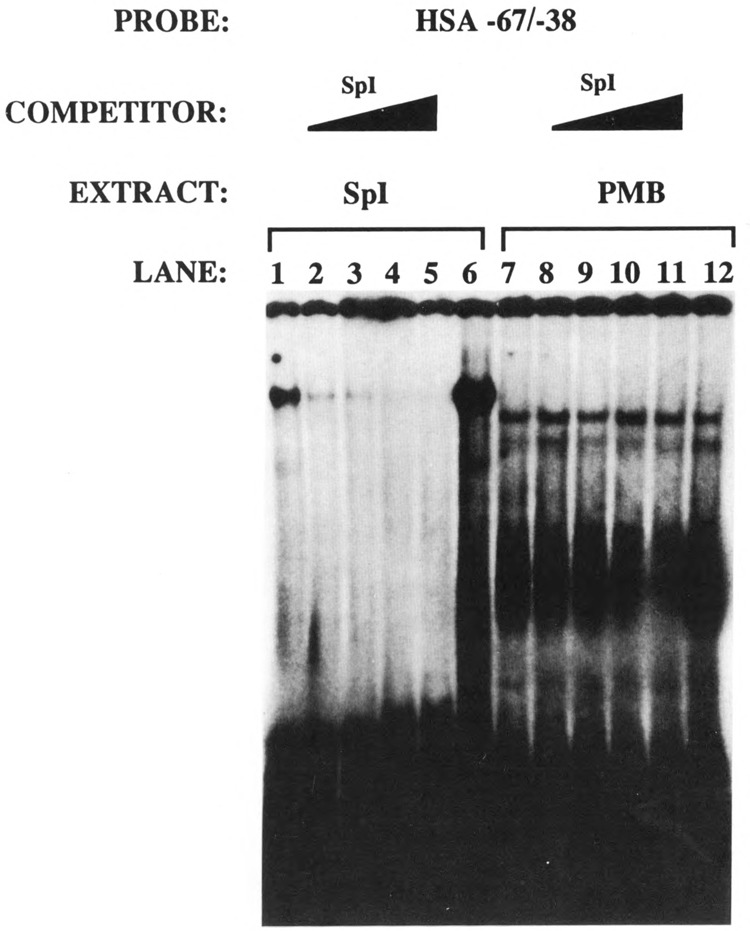
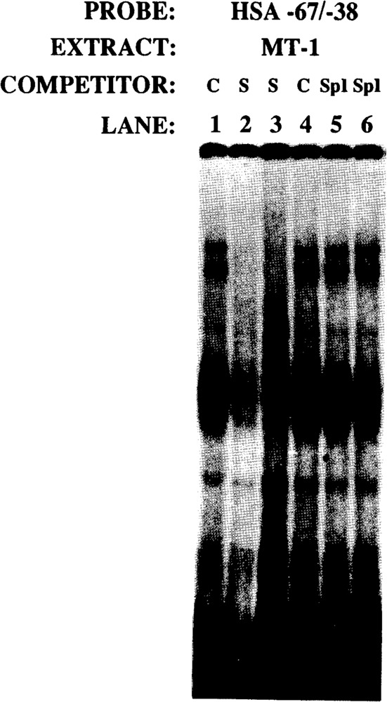
The skeletal alpha-actin gene is a member of the sarcomeric contractile protein gene family and is specifically expressed in differentiated muscle. The skeletal alpha-actin gene is regulated efficiently by enhancer and regulatory sequences between nucleotide positions -1282 and -87. In the present study we have shown that the sequences 3' of nucleotide position -87 can functionally interact with the SV40 enhancer in a tissue-specific manner and can restrict the ubiquitous function of the SV40 enhancer to myogenic cells. Site-specific cassette mutagenesis was used to delimit the sequences upstream of the TATA motif (-32), between nucleotide positions -64 and -37, that mediate efficient expression in myogenic cells in the presence of the SV40 enhancer. The skeletal alpha-actin promoter was trans-activated by the helix-loop-helix (HLH) transcription factors MyoD, MRF-4, and Myogenin, in pluripotential 10T1/2 fibroblasts and trans-repressed by the HLH protein Id (inhibitor of differentiation) in myogenic C2C12 cells. This trans-regulation required sequences upstream of -87 and occurred independently of the two consensus E boxes (CANNTG) at positions +18 and +71. The -64/-37 region interacted with purified Sp1 and an unidentified protein(s), proximal regulatory factor(s) I (PRF-I). We conclude that the muscle-specific expression of the skeletal alpha-actin promoter is not simply determined by MyoD elements and enhancer and regulatory sequences, but that the minimal promoter contains important determinants of cell-specific transcription that can function independently of the helix-loop-helix transcription factors.


Figures

Figure 1
Figure 1
A. Prototype recombinant plasmid pCAT-3′SV, used for the construction of the various promoter mutants. This vector contains a promoterless CAT gene with the SV40 enhancer situated at the 3′ end of the CAT reporter gene. B. The nucleotide sequence of the human skeletal α-actin gene between nucleotide positions −87 and +239. The TATA and E-box motifs are underlined.
Figure 2
Figure 2
Transient CAT assays in myogenic C2C12 cells demonstrating the cooperative interaction between the myogenic basal promoter sequences and the SV40 enhancer in muscle cells. Expression levels are shown as a percentage relative to pSV2CAT, set at 100%. The plasmids pCAT-3′SV, pHSA87CAT, pHSA87CAT-3′SV, pHSA2000CAT, pHCA47CAT, pHCA47CAT-3′SV, pHCA485CAT, and pSV2CAT are described in Materials and Methods.
Figure 3
Figure 3
Transient CAT assays in CV-l non-muscle cells demonstrating that the myogenic basal promoter sequences restrict the function of the SV40 enhancer to myogenic cells. Expression levels are shown as a percentage relative to pSV2CAT, set at 100%. The plasmids pCAT-3′SV, pHSA87-CAT pHSA87CAT-3′SV, pHSA-2000CAT, pHCA47CAT, pHCA-47CAT-3′SV, pHCA485CAT, and pSV2CAT are described in Materials and Methods.
Figure 4
Figure 4
Schematic representation of the cis-acting native human skeletal α-actin sequences and respective mutations cloned into the Xba I/Hind III cloning sites of the pCAT-3′SV vector.
Figure 5
Figure 5
Transient CAT assays demonstrating the effect of various deletions and mutations on the transcriptional interaction between myogenic sequences and the SV40 enhancer in muscle and non-muscle cells. Shown here are the levels of expression of plasmids in myogenic C2 and pluripotential 10T1/2 cells relative to the basal control vector pCAT-3′SV, arbitrarily set at 1. The plasmids pHSA87CAT-3′SV Ml, pHSA87CAT-3′SV M2, and pHSA87CAT-3′SV M3 have cassettes of 8 nucleotides mutated between nucleotide positions −87/−80, −79/−71, and −70/−63 respectively in the cis-acting region between nucleotide positions −87/+239 (shown in Figure 4). The plasmids pHSA37CAT-3′SV and pHSA37CAT-3′SV M4 contained the sequences between nucleotide positions −37/+239, with M4 harboring an SV40 TATA motif rather than the native myogenic TATA box. The plasmid pHSA87/30CAT-3′SV M1 contained the sequences between −87 and +30 carrying the M1 mutation into the pCAT-3′SV vector and did not contain the E box at nucleotide position +71. The plasmid pHSA87/30CAT-3′SV M1 M5 was identical to the former plasmid, except that the E box at position +18 was mutated, as shown in Figure 4. The plasmid pHSA37/30CAT-3′SV contained the sequences between −37 and +30 cloned into the pCAT-3′SV vector and did not contain the E box at nucleotide position +71. This plasmid was then modified to construct the plasmid pHSA37/30CAT-3′SV M5, which was identical to the former plasmid except that the E box at position +18 was mutated, as shown in Figure 4. The results shown represent the mean ± SD of 3–5 independent experiments.
Figure 6
Figure 6
Transient CAT assays in 10T1/2 cells demonstrating the ability of the myogenic specific helix-loop-helix proteins to transactivate the human skeletal α-actin promoter. Lanes 1–12 contained the plasmid pHSA2000CAT (6 μg), which included the full-length native human skeletal α-actin promoter. Lanes 3–6 also contained 0.5, 1, 2, and 4.0 μg of co-transfected pEMSV-MyoD. Lanes 7–10 also contained 0.5, 1, 2, and 4.0 μg of co-transfected pEMSV-myogenin. Lanes 11–14 also contained 0.5, 1, 2, and 4.0 μg of co-transfected pEMSV-MRE-4. The total amount of DNA in each transfection was kept at a constant 10 μg with the carrier plasmid, pUC18.
Figure 7
Figure 7
Transient CAT assays in C2C12 cells demonstrating the ability of the Id (inhibitor of differentiation) helix-loop-helix protein to trans-repress the human skeletal α-actin promoter. Lanes 1–8 contained the plasmid pHSA2000CAT (6 μg), which included the full-length native human skeletal α-actin promoter. Lanes 3–5 also contained 0.5, 1, and 2 μg of co-transfected pEMSV-E12(A.S.). Lanes 6–8 also contained 0.5, 1, and 2 μg of co-transfected pEMSV-Id. The total amount of DNA in each transfection was kept at a constant 10 μg with the carrier plasmid, pUC18.
Figure 8
Figure 8
Transient CAT assays in 10T1/2 cells demonstrating the effect of the myogenic specific helix-loop-helix proteins on deleted skeletal α-actin promoters. A. Lanes 1–10 contained the plasmid pHSA87CAT-3′SV M1 (6 μg); Lanes 3–6 also contained 0.5, 1, 2, and 4.0 μg of co-transfected pEMSV-MyoD. Lanes 7–10 also contained 0.5, 1, 2, and 4.0 μg of co-transfected pEMSV-myogenin. The total amount of DNA in each transfection was kept at a constant 10 μg with the carrier plasmid pUC18. B. Lanes 1–10 contained the plasmid pHSA37CAT-3′SV, and lanes 3–10 were transfected as shown in Figure 9A.
Figure 9
Figure 9
Transient CAT assays in C2C12 cells demonstrating the effect of the Id (inhibitor of differentiation) helix-loop-helix protein on deleted α-actin promoters. Lanes 1–8 of each assay contained the following plasmids: A, pHSA87CAT-3′SV M1; B, pHSA87CAT-3′SV M1,M5; C, pHSA37/30CAT-3′SV; and D, pHSA37/30CAT-3′SV M5 (6 μg). All plasmids included the full-length native human skeletal α-actin promoter. Lanes 3–5 also contained 0.5, 1, and 2 μg of co-transfected pEMSV-E12(A.S.). Lanes 6–8 also contained 0.5, 1, and 2 μg of co-transfected pEMSV-Id. The total amount of DNA in each transfection was kept at a constant 10 μg with the carrier plasmid pUC18.
Figure 10
Figure 10
The cis-acting region between nucleotide positions −67/−38 interacts with Spl and an unidentified factor, PRF-1, in myogenic nuclear extracts. The HSA −67/−38 probe was incubated with nuclear extracts derived from proliferating (PMB) and confluent (CMB) myoblasts and myotubes after 2 (MT-2) and 4 (MT-4) days of mitogen withdrawal (see Materials and Methods).
Figure 11
Figure 11
Spl and PRF-1 are distinct nuclear factors. The HSA −67/−38 probe was incubated with purified Spl (lanes 1–6) and PMB nuclear extract (lanes 7–12). Lanes 1, 6, 7, and 12 were control electrophorec tic mobility shift assays that did not contain any competitor DNAs. Lanes 2–5 and 8–11 contained 10-, 20-, 40-, and 80-fold molar excess of an Spl consensus sequence.
Figure 12
Figure 12
PRF-1 complex is specifically competed by HSA −67/−38, but not by Spl consensus sequences. The HSA −67/−38 probe was incubated with MT-1 nuclear extract; the control reactions are shown in lanes 1 and 4. Lanes 2 and 3, and 5 and 6, show competition with 40- and 80-fold molar excesses of self(S) and Spl consensus, respectively.

Similar articles

Cited by

References

    1. Bains W., Ponte P., Blau H., and Kedes L. (1984), Mol Cell Biol 4, 1449–1453. - PMC - PubMed
    1. Benezra R., Davis R. L., Lockshon D., Turner D. L., and Weintraub H. (1990), Cell 61, 49–59. - PubMed
    1. Blackwell T. K. and Weintraub H. (1990), Science 250, 1104–1110. - PubMed
    1. Bouvagnet P. F., Strehler E. E., White G. E., Strehler-Page M.-A., Nadal-Ginard B., and Mahdavi V. (1987), Mol Cell Biol 7, 4377–4389. - PMC - PubMed
    1. Boxer L. M., Prywes R., Roeder R. G., and Kedes L. (1989), Mol Cell Biol 9, 515–522. - PMC - PubMed

Publication types

MeSH terms

LinkOut - more resources