

Review
doi: 10.1016/0168-9525(92)90263-4.
Ordering gene function: the interpretation of epistasis in regulatory hierarchies
Affiliations
- PMID: 1365397
- PMCID: PMC3955268
- DOI: 10.1016/0168-9525(92)90263-4
Item in Clipboard
Review
Ordering gene function: the interpretation of epistasis in regulatory hierarchies
Trends Genet.
1992 Sep.
Abstract
The order of action of genes in a regulatory hierarchy that is governed by a signal can often be determined by the method of epistasis analysis, in which the phenotype of a double mutant is compared with that of single mutants. The epistatic mutation may be in either the upstream or the downstream gene, depending on the nature of the two mutations and the type of regulation. Nevertheless, when the regulatory hierarchy satisfies certain conditions, simple rules allow the position of the epistatic locus in the pathway to be determined without detailed knowledge of the nature of the mutations, the pathway, or the molecular mechanism of regulation.
Figures
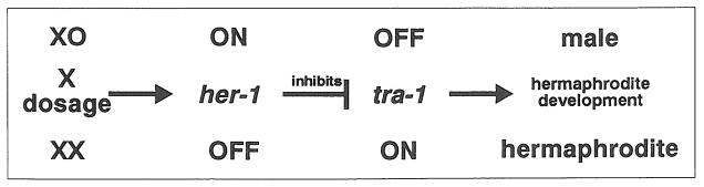
A model for sex determination in C. elegans. In C. elegans sex is determined by X chromosome dosage, acting through two genes her-1 and tra-1. When there is just one X chromosome (XO), her-1 is ON and inhibits tra-1, allowing male development. When there are two X chromosomes (XX), her-1 is OFF, allowing expression of tra-1, which causes hermaphrodite development.
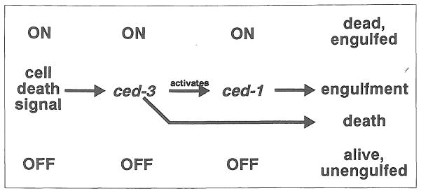
A model for the control of programmed cell death in C. elegans. In C. elegans development many cells undergo a stereotyped cell death program. The genes ced-1 and ced-3 are necessary for the normal completion of this program. The current model proposes that in cells fated to die a signal activates ced-3, causing cell death and turning on ced-1, which causes the dead cell to be engulfed by its neighbors. In a cell that would not normally die, ced-3 remains inactive, so the cell remains alive. Furthermore, because ced-3 is inactive, ced-1 also remains inactive, and the cell is not engulfed.
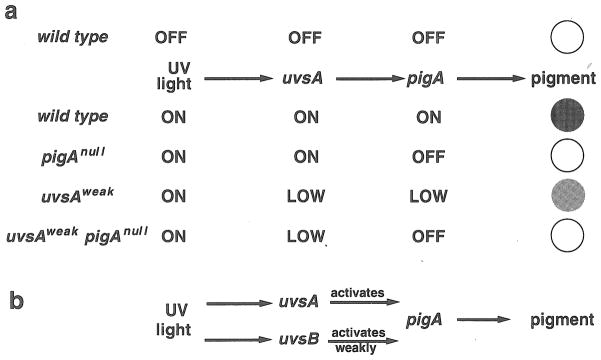
An example of misapplication of epistasis analysis. (a) In this hypothetical example a bacterium responds to UV light by synthesizing a protective pigment (shown as dark gray). The gene pigA, which encodes an enzyme that synthesizes the pigment, is activated in the presence of UV by a sensor encoded by uvsA. In the absence of UV, both genes are off and the bacteria produce a white colony. In the presence of UV both are fully on, and a pigmented colony results. A pigA null mutant (pigAnul1) is incapable of producing pigment and so remains white even in the presence of UV. A uvsA partial loss-of-function mutant (uvsAweak) responds only weakly to UV, producing low pigA activity and a slightly pigmented colony. The pigA null, although downstream, is epistatic to the uvsA partial loss-of-function mutation, since the double mutant remains white in UV. (b) In this variation of the model, UV light is sensed by two sensors, a major sensor encoded by uvsA and a minor sensor encoded by uvsB, which by itself can only weakly activate pigA. The epistasis relationship between a uvsA null mutation and a pigA null mutation in this model is identical to that between the uvsA partial loss-of-function mutation and the pigA null mutation in part (a). In particular, the uvsA null single mutant makes a slightly pigmented colony in UV light, because uvsB is still able to weakly activate pigA, and the double mutant makes a white colony, because it cannot synthesize pigment.
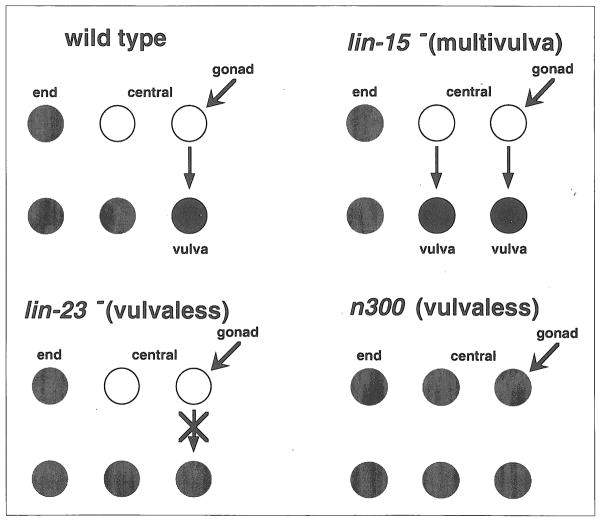
Vulval development in C. elegans. In wild-type C. elegans larvae, there are two types of epidermal blast cells. Those in the center of the worm (white) have the potential to form a vulva in response to a signal from the gonad. Those near the ends (gray) do not. In multivulva mutants such as lin-15, all central cells form vulvae, even if they don’t receive a signal from the gonad. In most vulvaless mutants (e.g. let-23) the central cells cannot perceive the gonadal signal and do not form a vulva. In an exceptional vulvaless mutant, n300, central cells take on the fates of end cells, so that there are no cells with the potential for vulval lineages.
References
Publication types
MeSH terms
Grants and funding
LinkOut - more resources
Full Text Sources
Other Literature Sources
