. 2003 Aug;9(4):745-56.
doi: 10.1089/107632703768247421.

Platelet-derived growth factor gene delivery stimulates ex vivo gingival repair

Affiliations

Platelet-derived growth factor gene delivery stimulates ex vivo gingival repair

Orasa Anusaksathien et al. Tissue Eng. 2003 Aug.

Abstract

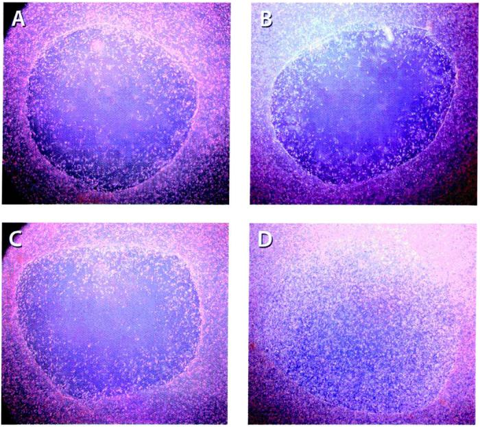
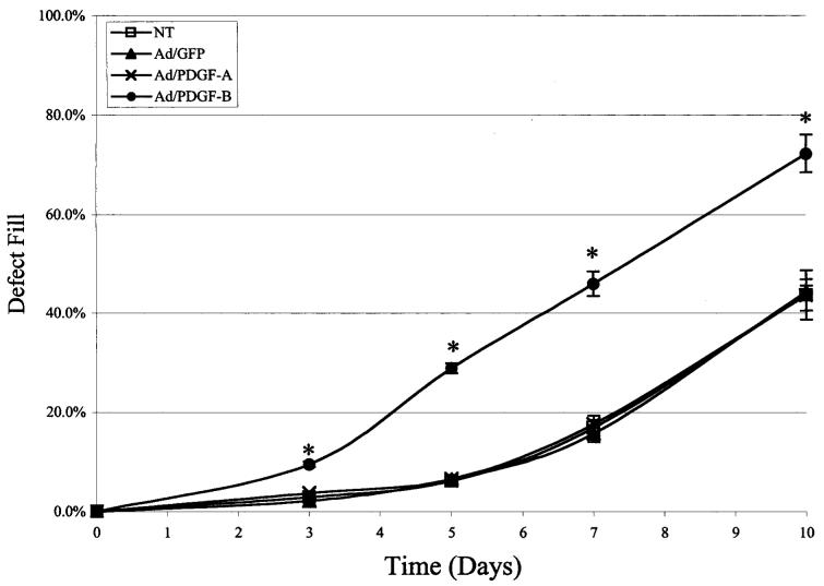
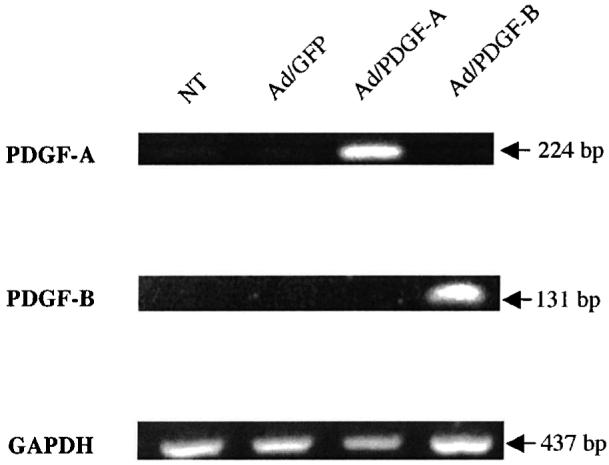
Destruction of tooth support due to the chronic inflammatory disease periodontitis is a major cause of tooth loss. There are limitations with available treatment options to tissue engineer soft tissue periodontal defects. The exogenous application of growth factors (GFs) such as platelet-derived growth factor (PDGF) has shown promise to enhance oral and periodontal tissue regeneration. However, the topical administration of GFs has not led to clinically significant improvements in tissue regeneration because of problems in maintaining therapeutic protein levels at the defect site. The utilization of PDGF gene transfer may circumvent many of the limitations with protein delivery to soft tissue wounds. The objective of this study was to test the effect of PDGF-A and PDGF-B gene transfer to human gingival fibroblasts (HGFs) on ex vivo repair in three-dimensional collagen lattices. HGFs were transduced with adenovirus encoding PDGF-A and PDGF-B genes. Defect fill of bilayer collagen gels was measured by image analysis of cell repopulation into the gingival defects. The modulation of gene expression at the defect site and periphery was measured by RT-PCR during a 10-day time course after gene delivery. The results demonstrated that PDGF-B gene transfer stimulated potent (>4-fold) increases in cell repopulation and defect fill above that of PDGF-A and corresponding controls. PDGF-A and PDGF-B gene expression was maintained for at least 10 days. PDGF gene transfer upregulated the expression of phosphatidylinosital 3-kinase and integrin alpha5 subunit at 5 days after adenovirus transduction. These results suggest that PDGF gene transfer has potential for periodontal soft tissue-engineering applications.


Figures

FIG. 1
FIG. 1
Fabrication of the bilayered wound defect model. (A) Fibroblast-populatedcollagen lattices (FPCLs) were fabricated and wounded with a 6-mm-diameter biopsy punch. Acellular collagen lattices were constructed and served as the inferior layer wound bed. Each FPCL was overlaid on the acellular layer followed by application of a collagen gel incorporated with 1% PPP (no treatment, NT) or 3.5 × 10⁶ plaque-forming units (PFU)/ml (20 MOI) of adenovirus encoding either GFP, PDGF-A, or PDGF-B. Images depict HGFs migrating from the defect margin. (B) A representative phase-contrast image depicts the wound margin of a defect treated with Ad/GFP for 7 days (original magnification, ×200). Arrows indicate HGF cells transduced by Ad/GFP near the defect margin. (C) Fluorescence image depicts the corresponding HGF cells expressing green fluorescent protein.
FIG. 2
FIG. 2
PDGF-B gene transfer stimulates ex vivo gingival defect fill. Images depict HGF repopulation into the wound defect area. Standardized digital images show HGFs filling in the wound area 10 days after no treatment (NT; A) or after treatment with 20 MOI of Ad/GFP (B), Ad/PDGF-A (C), or Ad/PDGF-B (D) per defect (n = 4 defects per group; original magnification, ×20).
FIG. 3
FIG. 3
PDGF-B gene transfer stimulates ex vivo gingival defect fill. HGF repopulation of created defects left untreated (NT) or treated with Ad/GFP, Ad/PDGF-A, or Ad/PDGF-B was measured by computer-assisted image analysis on days 3, 5, 7, and 10 postwounding. The percentage of wound fill was measured by determining the mean distance of cell migration into the wound area compared with the mean diameter of the wound. The data represent mean and standard error of measurement at each time point. Ad/PDGF-B induced significantly higher wound fill over time (*p < 0.001) compared with the other groups 3, 5, 7, and 10 days after gene transfer (n = 4 defects per group).
FIG. 4
FIG. 4
Density of cell repopulation into collagen wound defects. Total cell density of HGFs that repopulated defects left untreated (NT) or treated with Ad/GFP, Ad/PDGF-A, or Ad/PDGF-B was measured by computer-assisted image analysis on days 3, 5, 7, and 10. The data represent means ± SEM. Ad/PDGF-B induced significantly higher total cell density (*p , 0.001) compared with all other groups 3, 5, 7, and 10 days after gene transfer (n = 4 defects per group).
FIG. 5
FIG. 5
Prolonged induction of PDGF-A and PDGF-B transgenes in HGFs: day 10. RNAs extracted from HGF-populated lattices were subjected to RT-PCR. The PCR products were analyzed on ethidium bromide-stained gels and the expected PCR sizes are indicated. PDGF-A and PDGF-B genes were induced at all time points up to day 10 in the Ad/PDGF-A and Ad/PDGF-B groups, respectively.
FIG. 6
FIG. 6
Effect of PDGF-A and PDGF-B gene transfer on HGF gene expression by RT-PCR. RNAs extracted from HGF-populated lattices were subjected to RT-PCR with various sets of primers (Table 1) to determine the expression of ColIα1, PI3 kinase (P85α), and integrin α5 subunit (α5) compared with GAPDH (A) on day 5 and (B) on day 10. Histograms depict the relative ratio of gene expression normalized to GAPDH on day 5 (A, right) and day 10 (B, right). The PCR products were analyzed on ethidium bromide-stained gels and the expected PCR sizes are indicated. ColIα1 expression levels were not changed appreciably in response to Ad/PDGF-A and Ad/PDGF-B during the 10-day observation period. PI3 kinase (P85α) and integrin α5 subunit (α5) were upregulated in HGFs by PDGF-B induction on day 5, and remained unchanged compared with the other groups on day 10.

References

    1. Anusaksathien O, Giannobile WV. Growth factor delivery to re-engineer periodontal tissues. Curr. Pharm. Biotechnol. 2002;3:129. - PubMed
    1. Rutherford RB, Niekrash CE, Kennedy JE, Charette MF. Platelet-derivedand insulin-likegrowth factors stimulate regeneration of periodontal attachment in monkeys. J. Periodontal Res. 1992;27:285. - PubMed
    1. Giannobile WV, Finkelman RD, Lynch SE. Comparison of canine and nonhuman primate animal models for periodontal regenerative therapy: Results following a single administration of PDGF/IGF-I. J. Periodontol. 1994;65:1158. - PubMed
    1. Giannobile WV, Hernandez RA, Finkelman RD, Ryan S, Kiritsy CP, D'Andrea M, Lynch SE. Comparative effects of platelet-derived growth factor-BB and insulin-like growth factor-I, individually and in combination, on periodontal regeneration in Macaca fascicularis. J. Periodontal Res. 1996;31:301. - PubMed
    1. Howell TH, Fiorellini JP, Paquette DW, Offenbacher S, Giannobile WV, Lynch SE. A phase I/II clinical trial to evaluate a combination of recombinant human platelet-derived growth factor-BB and recombinant human insulin-like growth factor-I in patients with periodontal disease. J. Periodontol. 1997;68:1186. - PubMed

Publication types

Substances