. 2003 Sep;24(8):1560-9.

Diagnosing variant Creutzfeldt-Jakob disease with the pulvinar sign: MR imaging findings in 86 neuropathologically confirmed cases

Affiliations

Diagnosing variant Creutzfeldt-Jakob disease with the pulvinar sign: MR imaging findings in 86 neuropathologically confirmed cases

Donald A Collie et al. AJNR Am J Neuroradiol. 2003 Sep.

Abstract

Background and purpose: Variant Creutzfeldt-Jakob disease (vCJD) is a rare but important cause of dementia and death in young patients and is causally linked to bovine spongiform encephalopathy. Symmetrical hyperintensity in the pulvinar (posterior) nuclei of the thalamus (pulvinar sign) on brain MR images was described as a specific, noninvasive, diagnostic sign of vCJD in a previous small series. This purpose of this larger study was to evaluate this sign prospectively and further define the MR imaging characteristics of vCJD.

Methods: As part of the ongoing surveillance program in the United Kingdom, MR images of suspected cases of vCJD were collected during a 6-year period. All available images were assessed prospectively by one observer for the presence of the pulvinar sign. Images of neuropathologically confirmed cases were then assessed independently by two neuroradiologists for the degree of hyperintensity of the pulvinar on images of different MR sequences, and for the presence of abnormal hyperintensity in other areas of the brain. Discrepancies were reviewed jointly and a consensus opinion formed.

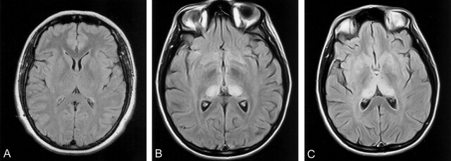
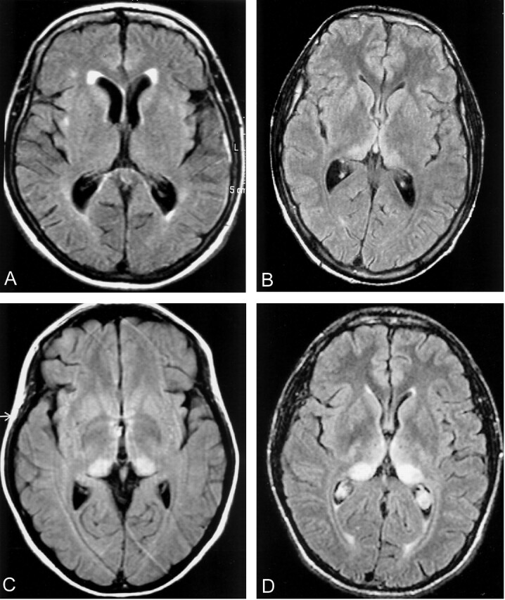
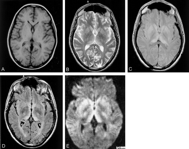
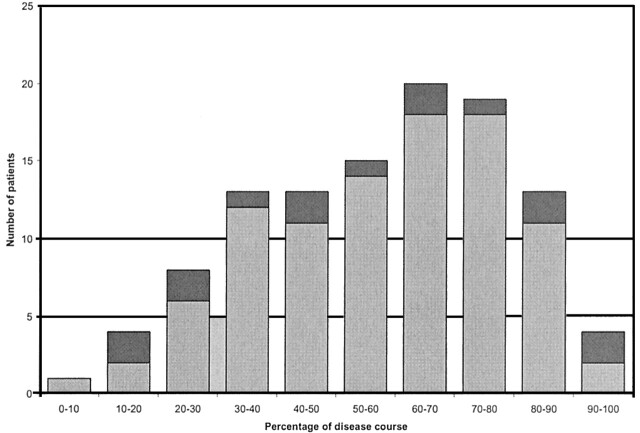
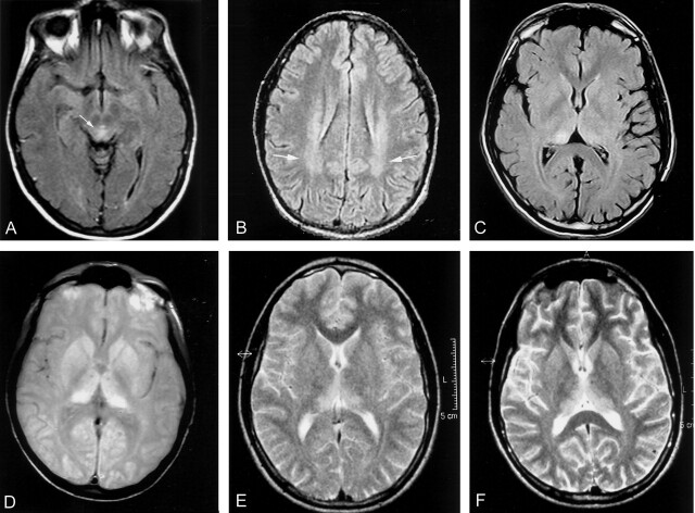
Results: Prospective analysis identified the pulvinar sign in 74 of 82 cases of vCJD. In the retrospective study, the pulvinar sign, as defined by hyperintensity of the pulvinar relative to the anterior putamen, was present on seven (9%) of 75 T1-weighted, 77 (71%) of 108 T2-weighted, 47 (81%) of 58 proton density-weighted, and 30 (100%) of 30 fluid-attenuated inversion-recovery (FLAIR) images. Diffusion-weighted images were available in two cases and were positive for the pulvinar sign in one. Other features were hyperintensity of the dorsomedial thalamic nuclei (93%), caudate head (40%), and periaqueductal gray matter (83%) on FLAIR images.

Conclusion: In the appropriate clinical context, demonstration of the pulvinar sign on MR images is a highly accurate diagnostic sign for vCJD. FLAIR sequence is more sensitive than other sequences. Positive MR images may obviate more invasive diagnostic tests in most cases.


Figures

F<sc>ig</sc> 1.
Fig 1.
A, Normal FLAIR image at the level of the basal ganglia shows the thalamus is normally isointense or slightly hypointense relative to the putamen. This appearance is depicted with most sequences, particularly the FLAIR sequence. B, Pulvinar sign of vCJD. FLAIR image shows marked, symmetrical hyperintensity of the pulvinar (posterior) thalamic nuclei. In this case, the pulvinar signal intensity was scored as grade 4 by both observers. C, “Hockey-stick” sign of vCJD. FLAIR image shows symmetrical pulvinar and dorsomedial thalamic nuclear hyperintensity. This combination gives a characteristic “hockey-stick” appearance and was present in 93% of cases with FLAIR imaging.
F<sc>ig</sc> 2.
Fig 2.
Grading of degree of pulvinar hyperintensity in vCJD. Because only hard-copy images were available in most cases of vCJD, a 5-point semiquantitative scoring system was used to grade the degree of hyperintensity of the cerebral structures, with white matter scored as 0, normal gray matter scored as 1, and pathologic hyperintensity scored from 2 to 4. Examples of FLAIR images are presented. A, Normal FLAIR image for comparison. B, Grade 2 hyperintensity of pulvinar. C, Grade 3 hyperintensity of pulvinar. D, Grade 4 hyperintensity of pulvinar
F<sc>ig</sc> 3.
Fig 3.
Effect of MR sequence on visibility of the pulvinar sign. A, Nonenhanced T1-weighted image. High signal intensity was relatively rarely detected on T1-weighted images, which was thought to be secondary to the T1-shortening effect from marked prion protein deposition. Contrast enhancement was not seen in any case. B–D, The pulvinar sign was more easily seen on T2-weighted (B) and PD-weighted (C) images, but was clearly visible on FLAIR (D) images. E, DW image was also positive in one of two available cases.
F<sc>ig</sc> 4.
Fig 4.
Graph shows timing of MR imaging in relation to vCJD duration. Dark gray indicates negative MR images; light gray, positive MR images
F<sc>ig</sc> 5.
Fig 5.
Other MR imaging findings in vCJD. A, Axial FLAIR image shows periaqueductal gray matter hyperintensity (arrow). Though not a specific sign, periaqueductal gray matter hyperintensity was seen in 83% of cases with FLAIR imaging. B, FLAIR image shows abnormal hyperintensity in the centrum semiovale white matter (arrows), reflecting the diffuse involvement of the brain by the disease. C, FLAIR image shows asymmetrical bilateral pulvinar hyperintensity. Asymmetrical hyperintensity was a rare finding in vCJD, seen in less than 5% of cases. D, PD-weighted image shows widespread basal ganglia hyperintensity. Though hyperintensity in the basal ganglia is also seen in a number of other conditions, the signal intensity of the pulvinar remains the most hyperintense, helping to categorize this case correctly as probable vCJD. E and F, Case of progressive cerebral atrophy in vCJD. Two images taken 3 months apart show subtle but definite global cerebral atrophy. Unlike in sCJD, cerebral atrophy is not a prominent feature of vCJD and is most easily seen in the parietooccipital region.

References

    1. Prusiner SB. Prions are novel infectious pathogens causing scrapie and Creutzfeldt-Jakob disease. Bioessays 1986;5:281–286 - PubMed
    1. Will RG, Ironside JW, Zeidler M, et al. A new variant of Creutzfeldt-Jakob disease in the UK. Lancet 1996;347:921–925 - PubMed
    1. Scott MR, Will R, Ironside J, et al. Compelling transgenetic evidence for transmission of bovine spongiform encephalopathy prions to humans. Proc Natl Acad Sci U S A 1999;96:15137–15142 - PMC - PubMed
    1. Prusiner SB. Prion diseases and the BSE crisis. Science 1997;278:245–251 - PubMed
    1. Will RG, Zeidler M, Stewart GE, et al. Diagnosis of new variant Creutzfeldt-Jakob disease. Ann Neurol 2000;47:575–582 - PubMed

Publication types