

Apparent diffusion coefficient measurements in the hippocampi in patients with temporal lobe seizures
- PMID: 13679274
- PMCID: PMC7973986
Apparent diffusion coefficient measurements in the hippocampi in patients with temporal lobe seizures
Abstract
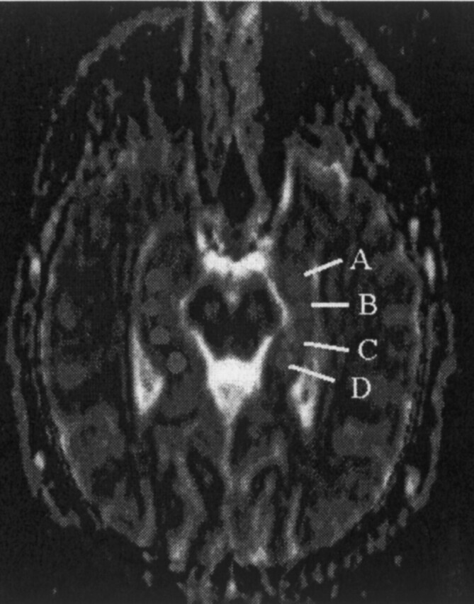
Background and purpose: Loss of neurons results in a relative increase in extracellular space that may lead to altered apparent diffusion coefficient (ADC) values in the hippocampi of patients with seizures. Our purpose was to determine if ADC values along the long axis of hippocampi are useful in evaluating patients with partial complex seizures.
Methods: Hippocampi of 23 patients with partial complex seizures and 25 healthy volunteers were evaluated with MR imaging and ADC maps. MR images were evaluated for loss of volume and/or high signal intensity on T2-weighted images and compared with ADC maps. ADCs were compared between patients and controls, as were ADCs along the length of each hippocampus. Mean and SDs were obtained for each measurement, and level of significance was determined (P <.05). The relationship between clinical lateralization and MR imaging and ADCs was studied.
Results: No significant variations were found in the ADCs in controls (side to side and along hippocampi). In patients, abnormalities were seen with MR imaging alone in 16, with ADC in 14, and with both in 21. Of 23 hippocampi with an abnormal MR appearance, 14 had abnormal ADCs. Nine hippocampi with a normal MR appearance had abnormal ADCs. Normal MR appearance and ADCs were seen in 13 hippocampi. Most abnormal ADCs were seen in the anterior aspect of the hippocampi. All differences were statistically significant. Of 19 patients who underwent clinical testing, unequivocal lateralization was established in 10. Concordance between clinical tests and MR imaging, ADC, and MR imaging plus ADC was found in five, five, and seven patients, respectively.
Conclusion: Visual assessment was better than ADCs alone for detection of abnormal hippocampi. MR imaging plus ADCs was better than either technique alone. ADCs may be abnormal when MR images are unremarkable. Concordance with clinical lateralization was better when MR imaging and ADC were jointly evaluated than when either technique was evaluated separately.
Figures
References
-
- Wetjen NM, Cohen-Gadol AA, Maher CO, et al. Frontal lobe epilepsy: diagnosis and surgical treatment. Neurosurg Rev 2002;25:119–138 - PubMed
-
- Chabolla D. Characteristics of the epilepsies. Mayo Clin Proc 2002;77:981–990 - PubMed
-
- Salanova V, Morris HH, Van Ness P, et al. Frontal lobe seizures: electroclinical syndromes. Epilepsia 1995;36:16–24 - PubMed
-
- Kramer U, Rivello JJ, Carmat P, et al. Clinical characteristics of complex partial seizures: a temporal versus a frontal lobe onset. Seizure 1997;6:57–61 - PubMed
-
- Van Paesschen W, Connelly A, Johnson Ch, Duncan JS. The amygdale and intractable temporal lobe epilepsy: a quantitative magnetic resonance imaging study. Neurology 1996;47:1021–1031 - PubMed
MeSH terms
LinkOut - more resources
Full Text Sources