

Delayed encephalopathy of acute carbon monoxide intoxication: diffusivity of cerebral white matter lesions
- PMID: 13679276
- PMCID: PMC7973971
Delayed encephalopathy of acute carbon monoxide intoxication: diffusivity of cerebral white matter lesions
Abstract
Background and purpose: Carbon monoxide intoxication has delayed effects on the cerebral white matter characterized by bilateral, confluent lesions that reflect diffuse demyelination. To increase our understanding of this process, we assessed the diffusion characteristics of these lesions.
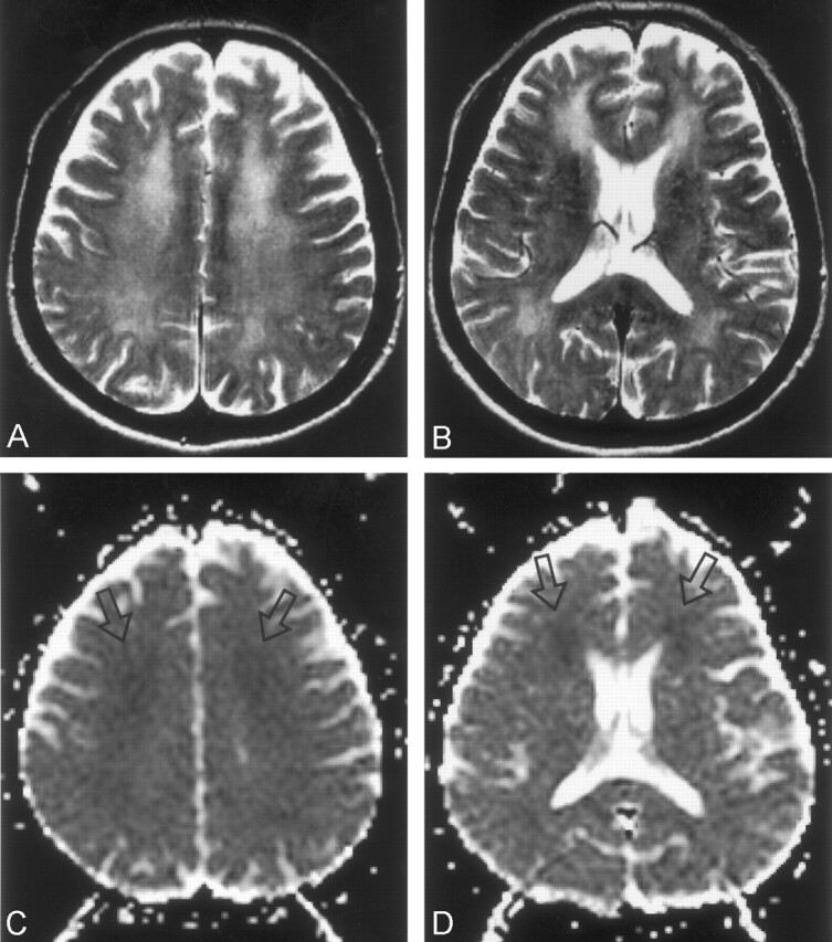
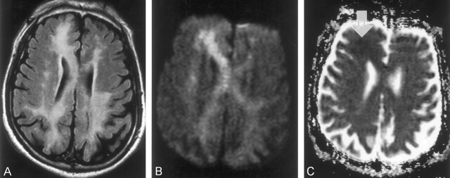
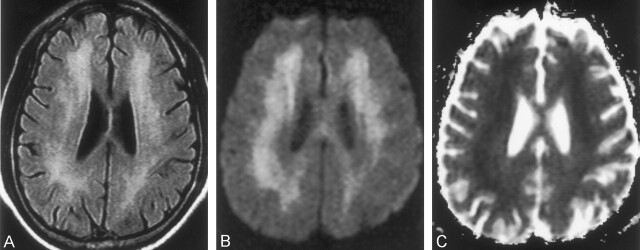
Methods: Five consecutive patients with delayed encephalopathy of CO intoxication were examined with diffusion MR imaging. Diffusion-weighted images (DWIs) were obtained 25-95 days after their exposure to CO and during a relapse of neuropsychiatric symptoms, which occurred after an initial recovery. Imaging was performed at 1.5 T by using a spin-echo echo-planar sequence with diffusion gradients of 0, 500, and 1000 s/mm(2). DWIs and apparent diffusion coefficient (ADC) maps were visually evaluated, and mean ADCs were calculated from the periventricular white matter and the centrum semiovale, where confluent hyperintensity was seen on T2-weighted images. Findings were compared with those of normal-looking white matter.
Results: In all five patients, both T2-weighted images and DWIs showed the white matter lesions as bilateral, diffuse, confluent areas of hyperintensity in the periventricular white matter and centrum semiovale. On ADC maps, these lesions were isointense, with focal areas of hypointensity (n = 4) or diffuse hypointensity (n = 1). Mean ADC values of the white matter lesions were significantly lower than those of normal-looking white matter, regardless of their isointensity or hypointensity on ADC maps (P <.05).
Conclusion: Bilateral, confluent, white matter lesions in patients with delayed encephalopathy of CO intoxication show decreased diffusivity.
Figures
References
-
- Choi IS. Delayed neurologic sequelae in carbon monoxide intoxication. Arch Neurol 1983;40:433–435 - PubMed
-
- Ginsberg MD. Delayed neurological deterioration following hypoxia. Adv Neurol 1979;26:21–44 - PubMed
-
- Roychowdhury S, Maljian JA, Galetta SL, Grossman RI. Postanoxic encephalopathy: diffusion MR findings. J Comput Assist Tomogr 1998;22:992–994 - PubMed
-
- Dooling EC, Richardson P Jr. Delayed encephalopathy after strangling. Arch Neurol 1976;33:196–199 - PubMed
-
- Ginsberg MD, Myers RE, McDonagh BF. Experimental carbon monoxide encephalopathy in the primate, II: clinical aspects, neuropathology, and physiologic correlation. Arch Neurol 1974;30:209–216 - PubMed
Publication types
MeSH terms
LinkOut - more resources
Full Text Sources
Medical