

Case Reports
Intralabyrinthine meningioma
Affiliations
- PMID: 13679286
- PMCID: PMC7974007
Item in Clipboard
Case Reports
Intralabyrinthine meningioma
AJNR Am J Neuroradiol.
2003 Sep.
Abstract
An 18-year-old female patient with unilateral hearing loss underwent evaluation with CT and MR imaging. A partially ossified, enhancing lesion in the bony labyrinth, with replacement of adjacent structures, was identified. Surgical biopsy revealed a meningioma arising primarily within the bony labyrinth. To our knowledge, this entity has not been previously described.
Figures
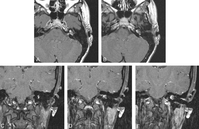
Dedicated thin-section, axial, T1-weighted (750/14/2/256 × 192/18 cm/2.5 mm/0.4 mm [TR/TE/NEX/matrix/FOV/section thickness/skip]) gadolinium-enhanced MR images show enhancement of the left labyrinthine lesion with minimal enhancement in the adjacent IAC.
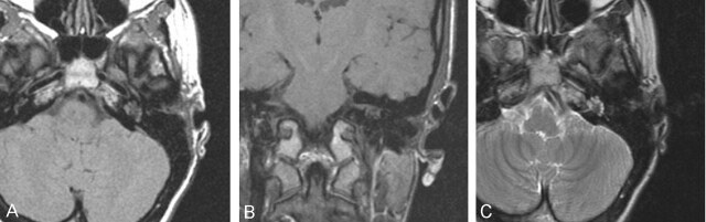
Dedicated thin-section axial (A) (750/14/2/256 × 192/18 cm/2.5 mm/0.4 mm [TR/TE/NEX/matrix/FOV/section thickness/skip]) and coronal (B) (600/14/2/256 × 192/18 cm/2.5 mm/0.4 mm) T1-weighted MR images obtained before gadolinium enhancement and axial fast spin-echo T2-weighted image (C) (5050/84/3/320 × 256/24 cm/5 mm/2.5 mm) demonstrate the labyrinthine mass, which is isointense to hypointense on both T1- and T2-weighted images.
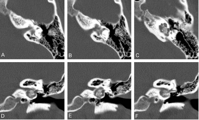
Direct axial (A–C) and coronal (D–F) 1-mm CT images of the left temporal bone obtained by using a bone algorithm. A partially ossified lesion, centered within the bony labyrinth, replaces the normal modiolus, vestibule, and basal turn of the cochlea. (1-mm section thickness, 512 × 512 matrix, 9.6-cm field of view, 9.6 cm/kV, 120/mAc 300).
References
-
- Nager GT. Meningiomas Involving the Temporal Bone. Springfield, IL: Charles C. Thomas;1964
-
- Guzowski J, Paparella MM, Nageswara K, Hoshino T. Meningiomas of the temporal bone. Laryngoscope 1976;86:1141–1146 - PubMed
-
- Luetje CM, Syms CA III, Luxford WE, et al. Meningiomas intrinsic to the geniculate ganglion. Am J Otol 1997;18:393–397 - PubMed
-
- Brookler KH, Hoffman RA, Camins M, Terzakis J. Trilobed meningioma: ampulla of posterior semicircular canal, internal auditory canal, and cerebellopontine angle. Am J Otol 1980;1:171–173 - PubMed
-
- Rinaldi A, Gazzeri G, Callovini GM, Masci P, Natali G. Acoustic intrameatal meningiomas. J Neurosurg Sci 2000;44:25–32 - PubMed
Publication types
MeSH terms
LinkOut - more resources
Full Text Sources
Medical