

Age-dependent cerebrovascular abnormalities and blood flow disturbances in APP23 mice modeling Alzheimer's disease
- PMID: 13679413
- PMCID: PMC6740368
- DOI: 10.1523/JNEUROSCI.23-24-08453.2003
Age-dependent cerebrovascular abnormalities and blood flow disturbances in APP23 mice modeling Alzheimer's disease
Abstract
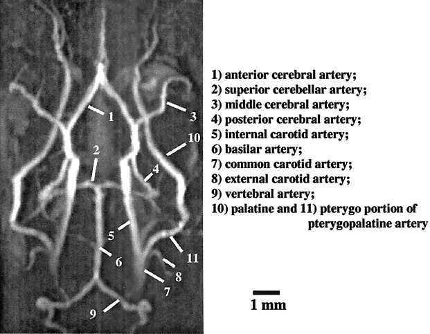
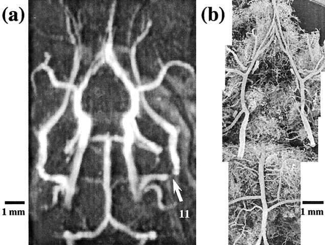
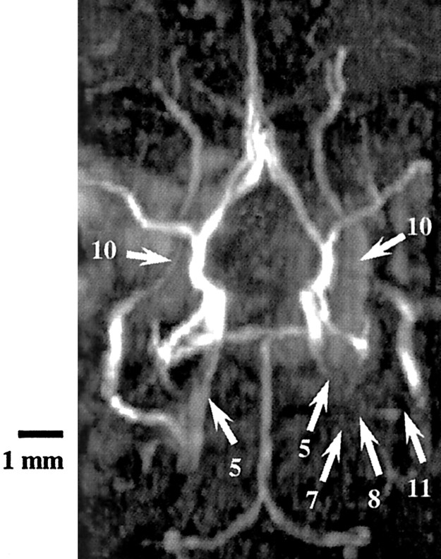
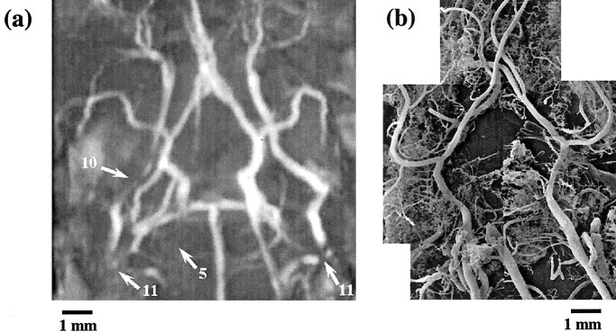
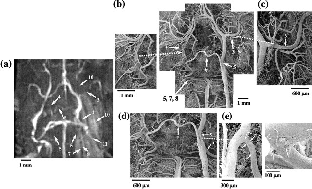
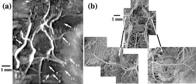
Neuropathological changes associated with Alzheimer's disease (AD) such as amyloidplaques, cerebral amyloid angiopathy, and related pathologies are reproduced in APP23 transgenic mice overexpressing amyloid precursor protein (APP) with the Swedish mutation. Magnetic resonance angiography (MRA) was applied to probe, in vivo, the cerebral arterial hemodynamics of these mice. Flow voids were detected at the internal carotid artery of 11-month-old APP23 mice. At the age of 20 months, additional flow disturbances were observed in large arteries at the circle of Willis. Vascular corrosion casts obtained from the same mice revealed that vessel elimination, deformation, or both had taken place at the sites where flow voids were detected by MRA. The detailed three-dimensional architecture of the vasculature visible in the casts assisted the identification of smaller vessels most likely formed as substitution or anastomosis within the circle of Willis. Angiograms and corrosion casts from nontransgenic, age-matched mice manifested no major abnormalities in the cerebrovascular arterial flow pattern. Because no transgene overexpression has been found in the cerebrovasculature of APP23 mice and no deposits of amyloid-beta (Abeta) were observed in large arteries in the region of the circle of Willis, the present results suggest that soluble Abeta may exert deleterious effects on the vasculature. Our findings support the idea that cerebral circulatory abnormalities evolving progressively could contribute to AD pathogenesis. The study also shows the power of MRA to identify changes of vascular function in genetically engineered mice. MRA as a noninvasive technique could be applied to test new therapeutic concepts in animal models of AD and in humans.
Figures
References
-
- Alsop DC, Detre JA, Grossman M ( 2000) Assessment of cerebral blood flow in Alzheimer's disease by spin-labeled magnetic resonance imaging. Ann Neurol 47: 93-100. - PubMed
-
- Andersen JK ( 2001) Genetically engineered mice and their use in aging research. Mol Biotechnol 19: 45-57. - PubMed
-
- Arendash GW, Su GC, Crawford FC, Bjugstad KB, Mullan M ( 1999) Intravascular beta-amyloid infusion increases blood pressure: implications for a vasoactive role of beta-amyloid in the pathogenesis of Alzheimer's disease. Neurosci Lett 268: 17-20. - PubMed
-
- Beckmann N ( 2000) High resolution magnetic resonance angiography noninvasively reveals mouse strain differences in the cerebrovascular anatomy in vivo. Magn Reson Med 44: 252-258. - PubMed
-
- Beckmann N, Stirnimann R, Bochelen D ( 1999) High resolution magnetic resonance angiography of the mouse brain: application to murine focal cerebral ischemia models. J Magn Reson 140: 442-450. - PubMed
Publication types
MeSH terms
Substances
LinkOut - more resources
Full Text Sources
Other Literature Sources
Medical
Molecular Biology Databases