. 2004 Jan;15(1):121-31.
doi: 10.1091/mbc.e03-05-0342. Epub 2003 Sep 17.

The Drosophila kinesin-like protein KLP67A is essential for mitotic and male meiotic spindle assembly

Affiliations

The Drosophila kinesin-like protein KLP67A is essential for mitotic and male meiotic spindle assembly

Rita Gandhi et al. Mol Biol Cell. 2004 Jan.

Abstract

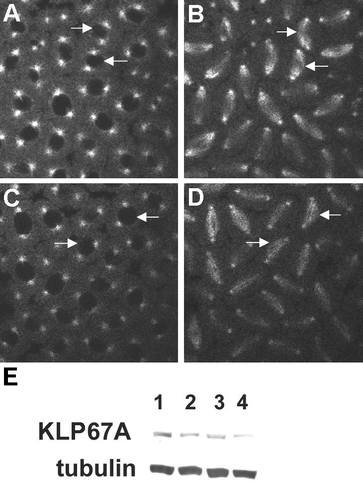
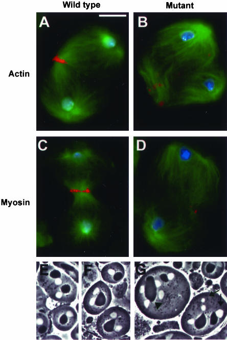
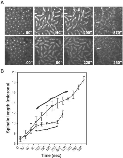
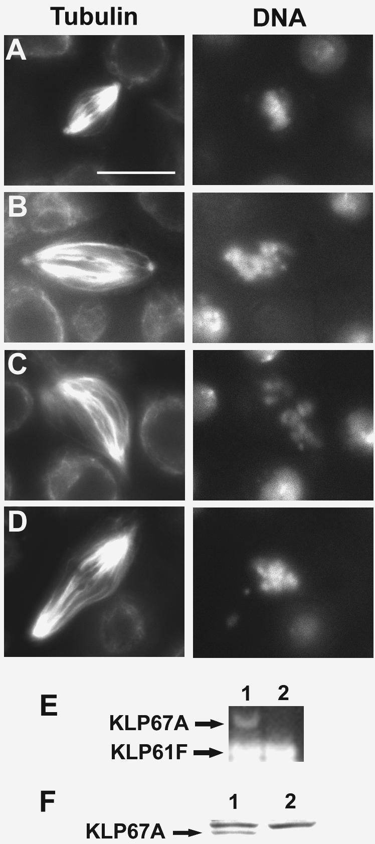
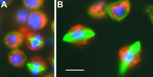
We have performed a mutational analysis together with RNA interference to determine the role of the kinesin-like protein KLP67A in Drosophila cell division. During both mitosis and male meiosis, Klp67A mutations cause an increase in MT length and disrupt discrete aspects of spindle assembly, as well as cytokinesis. Mutant cells exhibit greatly enlarged metaphase spindle as a result of excessive MT polymerization. The analysis of both living and fixed cells also shows perturbations in centrosome separation, chromosome segregation, and central spindle assembly. These data demonstrate that the MT plus end-directed motor KLP67A is essential for spindle assembly during mitosis and male meiosis and suggest that the regulation of MT plus-end polymerization is a key determinant of spindle architecture throughout cell division.


Figures

Figure 4.
Figure 4.
Klp67A mutations cause an incomplete centrosome separation. (A–D) Still images from real-time analysis of mitosis in blastoderm embryos of Klp67A322b24/Df(3L)29A6 (A and B) and wild-type (C and D) mothers. Note the correlation between incomplete centrosome separation and curved banana-shaped spindles (arrows). Incomplete centrosome separation is sometimes seen in wild-type (arrows in C), which also results in curved spindle (arrows in D) but is less frequent than in the mutant. (E) Western blot analysis of KLP67A expression. Protein homogenates were prepared from 0 to 2.5 h egg collections from mutant and wild-type mothers. The blot was probed with a rabbit polyclonal antibody to KLP67A as well as an antibody to α-tubulin. α-Tubulin was used as a loading control. 1, wild-type control. 2, Df(3L)29A6/TM6B. 3, Klp67A322b24/TM6B. 4, Klp67A322b24/Df(3L)29A6.
Figure 1.
Figure 1.
Klp67A is required for aster migration to the opposite sides of spermatocyte nuclei. Wild-type (A) and Klp67A322b24/Df(3L)29A6 (B–D) Primary spermatocyte preparations were stained for α-tubulin (green), centrosomin (red), and DNA (blue). (A) Prometaphase I spermatocyte showing two well separated prominent asters closely apposed to the nuclear envelope. (B) Two mutant prometaphase I spermatocytes with ectopically located asters, not associated with the nuclear envelope and still close to each other. (C) A mutant primary spermatocyte showing the bulk of one of the centrosomes and its associated aster detached from the spindle pole (arrowhead). (D) Tetraploid mutant spermatocyte with four ectopically located asters, indicating a cytokinesis failure in a previous gonial division.
Figure 2.
Figure 2.
Klp67A mutations affect spindle morphology of Drosophila spermatocytes. Wild-type (A, B, D, and F) and Klp67A322b24/Df(3L)29A6 (C, E, and G) meiotic cells were stained for α-tubulin (green), centrosomin (orange), and DNA (blue). (A) Wild-type metaphase I. (B) Wild-type anaphase I. (C) A mutant metaphase (arrowhead) with a bipolar spindle and an anaphase figure (arrow) showing an abnormal chromosome segregation. In both cells, the MTs seem to be longer than in their wild-type counterparts. (D) Two wild-type telophases I. (E) Three mutant telophases I showing abnormally long astral MTs and defective central spindles. (F) Two wild-type telophases II. (G) Mutant telophases II. The arrowhead points to a morphologically abnormal telophase figure in which interpolar MTs overlap in the middle of the cell but do not form a typical dense central spindle. The arrow points to two telophases within the same cytoplasm showing long astral MTs. Note the overlapping of astral MTs emanating from different spindles. Bar, 10 μm.
Figure 3.
Figure 3.
Klp67A mutations disrupt central spindle and contractile ring assembly and cause failures in both cytokinesis and chromosome segregation. Primary spermatocytes from wild-type (A and C) and Klp67A322b24/Df(3L)29A6 (B and D) males. Preparations were stained for α-tubulin (green), actin (A and B; red) or myosin (C and D; red) and DNA (blue). (A and C) Wild-type telophase I spermatocytes showing a prominent central spindle and a regular actomyosin ring. (B and D) Mutant telophase I figures showing a severely defective central spindle and irregular patches of either actin (b) or myosin (d) at the cleavage furrow. (E–G) Phase contrast images of wild-type (e) and Klp67A322b24/Df(3L)29A6 (F and G) living spermatids. (E) Wild-type partial cyst in which each spermatid consists of a phase-dark nebenkern and a phase-light nucleus. (F) Klp67A mutant spermatids consisting of two equally sized nuclei associated with a large nebenkern (G) A Klp67A spermatid consisting of a large nebenkern associated with four nuclei of normal size (arrowhead) and two spermatids showing irregularly sized nebenkern associated with micro- and macronuclei (arrows). Bar, 10 μm.
Figure 5.
Figure 5.
Klp67A mutations result in abnormally long and curved spindles and a failure of central spindle assembly. (A) Mutant embryos (Klp67A322b24/Df(3L)29A6; top four panels) and wild-type embryos [Df(3L)29A6/TM6B; bottom four panels] were injected with rhodamine-tubulin, and mitotic divisions were examined by time-lapse confocal microscopy. Panels show representative images of different mitotic stages; timing is indicated at the bottom of each panel. Note that the mutant spindles are abnormally long and curved and that midzone MTs are not apparent during anaphase B in the mutant as they are in wild type (arrows). (B) Plot of spindle length versus time beginning with nuclear envelope breakdown up until central spindle formation at the end of anaphase B of cycle 11. Note that mutant spindles are consistently longer that their wild-type counterparts and that there is a significant lengthening of the time spent in metaphase (compare bracketed intervals). Bar, SD; closed circles, wild-type; empty squares, mutant.
Figure 6.
Figure 6.
Klp67A dsRNA treatment in DL2 cells results in elongated spindles and mitotic arrest. (A–D) Cytological phenotypes caused by KLP67A depletion. Klp67A (RNAi) cells and control cells were fixed and stained for α-tubulin and DNA (with DAPI). (A) Mock-transfected control cells. (B–D) Cells treated with Klp67A dsRNA. (A) Metaphase. (B–D) Metaphase-like spindles in RNAi cells. Bar, 10 μm. (E) Semiquantitative RT-PCR assay shows undetectable levels of Klp67A mRNA in dsRNA-treated samples. 1, control; 2, cells treated with Klp67A dsRNA. Klp61F mRNA amplified by RT-PCR was used as an internal control. (F) Western blot analysis of DL2 cells treated with Klp67A dsRNA. Forty micrograms of protein was loaded in each lane, and the blot was probed with a rabbit polyclonal antibody to KLP67A. The common background band serves as a loading control.
Figure 7.
Figure 7.
KLP67A depletion affects the mitochondria distribution pattern in DL2 cells. Klp67A (RNAi) cells were fixed and stained with anti-α-tubulin (green), anti-F1ATP synthase (red) to stain mitochondria, and DAPI (blue). (A) Mock-transfected control cells. (B) Cells treated with Klp67A dsRNA. Bar, 10 μm.
Figure 8.
Figure 8.
Depletion of both KLP61F and KLP67A arrests cells in mitosis with monopolar spindles. DL2 cells were transfected with Klp67A dsRNA and Klp61F dsRNA, either separately or in combination. Cells were fixed and stained for α-tubulin and DNA (with DAPI). Note the monopolar spindle morphology in cells treated with either Klp61F dsRNA alone or with both Klp67A and Klp61F dsRNA. In the latter case, MTs are much longer than in the former. Bar, 10 μm.

References

    1. Adams, R.R., Tavares, A.A., Salzberg, A., Bellen, H.J., and Glover, D.M. (1998). pavarotti encodes a kinesin-like protein required to organize the central spindle and contractile ring for cytokinesis. Genes Dev. 12, 1483–1494. - PMC - PubMed
    1. Basto, R., Gomes, R., and Karess, R.E. (2000). Rough Deal and Zw10 are required for the metaphase checkpoint in Drosophila. Nat. Cell Biol. 2, 939–943. - PubMed
    1. Bonaccorsi, S., Giansanti, M.G., and Gatti, M. (1998). Spindle self-organization and cytokinesis during male meiosis in asterless mutants of Drosophila melanogaster. J. Cell Biol. 142, 751–761. - PMC - PubMed
    1. Cassimeris, L. (1999). Accessory protein regulation of microtubule dynamics throughout the cell cycle. Curr. Opin. Cell Biol. 11, 134–141. - PubMed
    1. Cenci, G., Bonaccorsi, S., Pisano, C., Verni, F., and Gatti, M. (1994). Chromatin and microtubule organization during premeiotic meiotic, and early postmeiotic stages of Drosophila melanogaster spermatogenesis. J. Cell Sci. 107, 3521–3534. - PubMed

Publication types

MeSH terms

LinkOut - more resources