. 2003 Sep 30;100(20):11406-11.
doi: 10.1073/pnas.1936664100. Epub 2003 Sep 17.

Beta-arrestin-mediated activation of MAPK by inverse agonists reveals distinct active conformations for G protein-coupled receptors

Affiliations

Beta-arrestin-mediated activation of MAPK by inverse agonists reveals distinct active conformations for G protein-coupled receptors

Mounia Azzi et al. Proc Natl Acad Sci U S A. .

Abstract

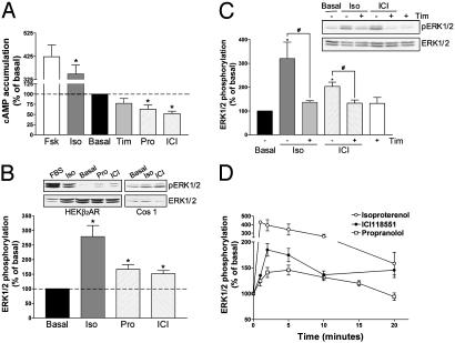
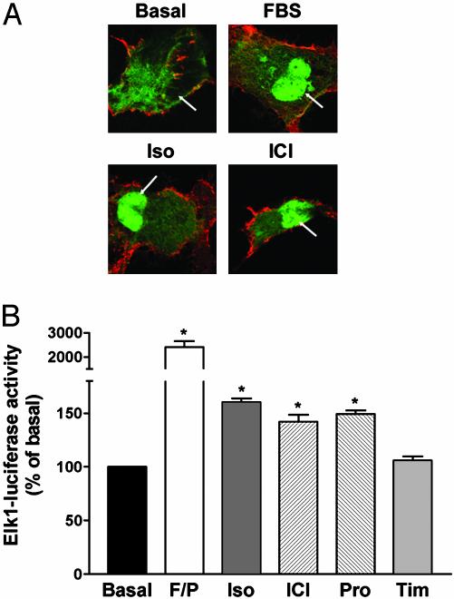
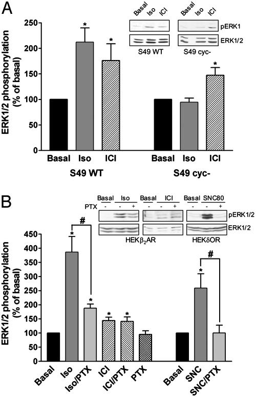
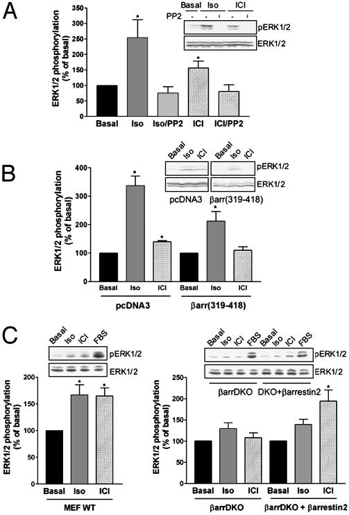
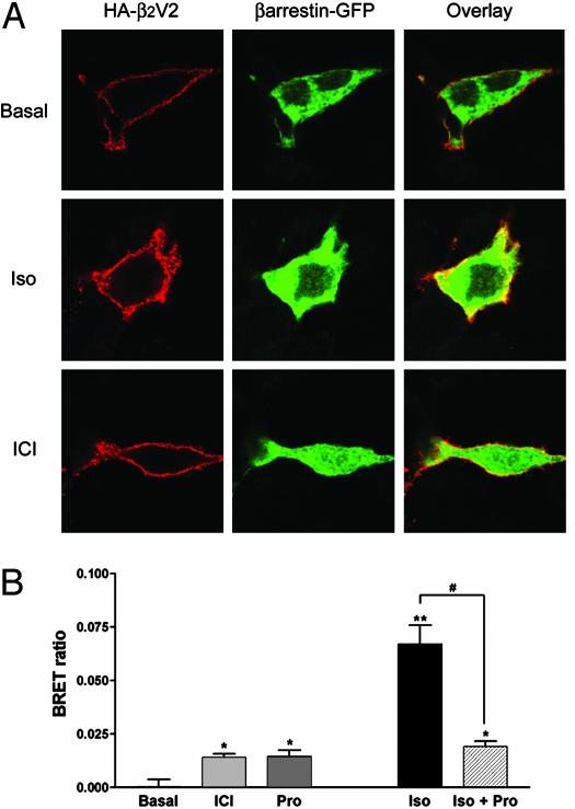
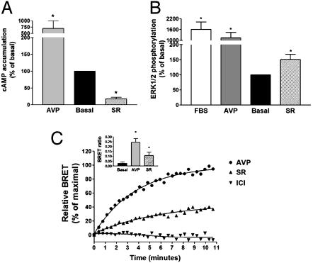
It is becoming increasingly clear that signaling via G protein-coupled receptors is a diverse phenomenon involving receptor interaction with a variety of signaling partners. Despite this diversity, receptor ligands are commonly classified only according to their ability to modify G protein-dependent signaling. Here we show that beta2AR ligands like ICI118551 and propranolol, which are inverse agonists for Gs-stimulated adenylyl cyclase, induce partial agonist responses for the mitogen-activated protein kinases extracellular signal-regulated kinase (ERK) 1/2 thus behaving as dual efficacy ligands. ERK1/2 activation by dual efficacy ligands was not affected by ADP-ribosylation of Galphai and could be observed in S49-cyc- cells lacking Galphas indicating that, unlike the conventional agonist isoproterenol, these drugs induce ERK1/2 activation in a Gs/i-independent manner. In contrast, this activation was inhibited by a dominant negative mutant of beta-arrestin and was abolished in mouse embryonic fibroblasts lacking beta-arrestin 1 and 2. The role of beta-arrestin was further confirmed by showing that transfection of beta-arrestin 2 in these knockout cells restored ICI118551 promoted ERK1/2 activation. ICI118551 and propranolol also promoted beta-arrestin recruitment to the receptor. Taken together, these observations suggest that beta-arrestin recruitment is not an exclusive property of agonists, and that ligands classically classified as inverse agonists rely exclusively on beta-arrestin for their positive signaling activity. This phenomenon is not unique to beta2-adrenergic ligands because SR121463B, an inverse agonist on the V2 vasopressin receptor-stimulated adenylyl cyclase, recruited beta-arrestin and stimulated ERK1/2. These results point to a multistate model of receptor activation in which ligand-specific conformations are capable of differentially activating distinct signaling partners.


Figures

Fig. 1.
Fig. 1.
Signaling efficacy of β2AR ligands. HEK293 cells stably expressing the β2AR (HEKβ2AR) were treated with the indicated drugs (1 μM), and cAMP accumulation (A) and ERK1/2 phosphorylation (B) were assessed after a 15-min and a 5-min incubation, respectively. Results are expressed as percentage of the value obtained in nonstimulated cells and represent the mean ± SEM of three to eight independent experiments. *, P < 0.01 compared with basal. (B Inset) Representative examples of ERK1/2 phosphorylation experiments carried out in HEKβ2AR and Cos1 cells. (C) Competitive antagonism of timolol (10 μM) on isoproterenol-mediated (10 nM) and ICI118551-mediated (10 nM) ERK1/2 phosphorylation (n = 8–10; *, P < 0.05 compared with basal cells; #, P < 0.05 for the indicated comparison). (D) Kinetics of ERK1/2 activation by different β2AR ligands (1 μM) in HEKβ2AR (circles) and canine cardiomyocytes (squares) (n = 4–7).
Fig. 2.
Fig. 2.
Nuclear translocation of activated ERK. (A) Subcellular redistribution of ERK2-GFP after FBS, isoproterenol (Iso) and ICI118551 (ICI) treatments. Cos1 cells transiently expressing HA-β2AR and ERK2-GFP were stimulated or not for 20 min with 10% FBS or 10 μM Iso or ICI. Arrows indicate the nucleus. Under basal conditions, 13% of the cells showed colocalization of ERK2-GFP with the nuclear marker To-Pro-3 iodine. After drug treatments, this value increased to 84% for FBS, 72% for Iso, and 67% for ICI. (B) Gene reporter assay for Elk-regulated transcription. Luciferase activity was measured after a 6-h incubation with 10 μM Iso, ICI, propranolol (Pro), or timolol (Tim). Stimulation with 10% FBS and 1 μM phorbol 12-myristate 13-acetate (F/P) was used as a positive control. Data are expressed as percentage of the value obtained in nonstimulated cells and represent the mean ± SEM of four to six independent experiments; *, P < 0.01 compared with basal.
Fig. 3.
Fig. 3.
Role of Gs and Gi in isoproterenol and ICI118551-mediated ERK1/2 activation. ERK1/2 phosphorylation was assessed in S49 wild-type (S49 WT) and S49 cyccells endogenously expressing the β2AR (A) or in HEK293 cells stably transfected with the β2AR (HEKβ2AR) and treated or not with 100 ng/ml pertussis toxin (PTX) for 16 h (B). Cells were stimulated with 1 μM isoproterenol (Iso) or ICI118551 (ICI) for 5 min. The efficacy of PTX pretreatment was controlled by its ability to block ERK1/2 activation in HEK293 cells expressing the Gi/o-coupled δ-opioid receptor (HEKδOR) by the δOR agonist SNC80 (1 μM; 5 min). Results are expressed as percentage of the value obtained in nonstimulated cells and represent the mean ± SEM of three to eight independent experiments. *, P < 0.05 compared with basal; #, P < 0.05 for the indicated comparison. Insets show representative experiments.
Fig. 4.
Fig. 4.
Involvement of Src and β-arrestin in isoproterenol- and ICI118551-mediated ERK1/2 activation. ERK1/2 phosphorylation was assessed in HEK293 cells stably expressing the β2AR pretreated or not with the cell-permeable Src tyrosine kinase inhibitor, PP2 (50 μM, 1 h) (A), or transiently transfected with the β-arrestin dominant negative βarr(319–418) or the empty vector pcDNA3 as a control (B). (C) ERK1/2 phosphorylation was assessed in mouse embryonic fibroblasts (MEF) derived from wild-type mice (WT) and from KO mice lacking both β-arrestin 1 and 2 (βarrDKO) that were transiently transfected with the human HA-β2AR. Expression levels of 150–250 fmol of β2AR per mg of protein were reached in both WT and KO cells. For rescue experiments, βarrDKO MEF were cotransfected with HA-β2AR and β-arrestin 2 (βarrDKO + β-arrestin 2). In all cases, cells were stimulated for 5 min with 1 μM isoproterenol (Iso) or ICI118551 (ICI). Data are expressed as percentage of the value obtained in nonstimulated cells and represent the mean ± SEM of three to eight independent experiments. *, P < 0.05 compared with basal. Insets show representative experiments.
Fig. 5.
Fig. 5.
β-arrestin recruitment. (A) Cos1 cells transiently expressing the HA-tagged β2V2R chimera and β-arrestin-2-GFP were stimulated or not for 15 min with 10 μM isoproterenol (Iso) or ICI118551 (ICI). Subcellular distribution was then assessed by fluorescence microscopy using Texas red-conjugated antibodies for the HA-β2V2R and direct fluorescence for the β-arrestin-GFP. (B) BRET was determined in HEK293 cells transiently coexpressing β2AR-GFP and β-arrestin-Rluc after incubation with 1 μM isoproterenol (Iso), ICI118551 (ICI), or propranolol (Pro) for 1 h at room temperature. Competitive antagonism of Pro on Iso-mediated β-arrestin recruitment was measured after pretreatment of the cells with 10 μM Pro for 15 min before stimulation with 0.1 μM Iso. Data are expressed as BRET ratio and represent the mean ± SEM of four independent experiments. *, P < 0.05 compared with nonstimulated cells (basal); **, P < 0.01 compared with nonstimulated cells (basal); #, P < 0.001 for the indicated comparison.
Fig. 6.
Fig. 6.
MAPK activation and β-arrestin recruitment by the V2R ligand SR121463B. HEK293 cells stably expressing the V2R were treated with 1 μM Arg-vasopressin (AVP), SR121463B (SR), or 10% FBS, and cAMP accumulation (A) and ERK1/2 phosphorylation (B) were assessed after 15 and 5 min of incubation, respectively. Results are expressed as percentage of the value obtained in nonstimulated cells and represent the mean ± SEM of three to four independent experiments. *, P < 0.05 compared with basal. (C) BRET was determined in HEK293 cells transiently coexpressing V2R-GFP and β-arrestin-Rluc in the presence of 1 μM AVP (•), SR (▴), or ICI (▾) for the indicated times. Data are expressed as percentage of the maximal BRET signal promoted by AVP and are representative of two to four independent experiments. (Inset) BRET ratio after a 5-min stimulation with 1 μM AVP or SR. Data represent the mean ± SEM of four independent experiments. *, P < 0.05 compared with nonstimulated cells (basal).

Similar articles

Cited by

References

    1. Gutkind, J. S. (2000) Sci. STKE 40, RE1. - PubMed
    1. Schmitt, J. M. & Stork, P. J. (2000) J. Biol. Chem. 275, 25342–25350. - PubMed
    1. Luttrell, L. M., Ferguson, S. S., Daaka, Y., Miller, W. E., Maudsley, S., Della Rocca, G. J., Lin, F., Kawakatsu, H., Owada, K., Luttrell, D. K., et al. (1999) Science 283, 655–661. - PubMed
    1. DeFea, K. A., Zalevsky, J., Thoma, M. S., Dery, O., Mullins, R. D. & Bunnett, N. W. (2000) J. Cell. Biol. 148, 1267–1281. - PMC - PubMed
    1. McDonald, P. H., Chow, C. W., Miller, W. E., Laporte, S. A., Field, M. E., Lin, F. T., Davis, R. J. & Lefkowitz, R. J. (2000) Science 290, 1574–1577. - PubMed

Publication types