. 2003 Sep 30;100(20):11285-90.
doi: 10.1073/pnas.1934283100. Epub 2003 Sep 17.

Insulin-induced phosphorylation of FKHR (Foxo1) targets to proteasomal degradation

Affiliations

Insulin-induced phosphorylation of FKHR (Foxo1) targets to proteasomal degradation

Hitomi Matsuzaki et al. Proc Natl Acad Sci U S A. .

Abstract

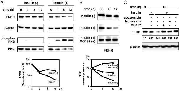
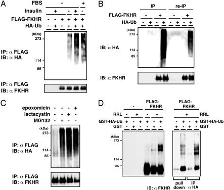
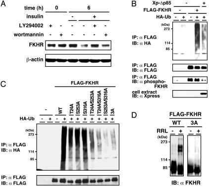
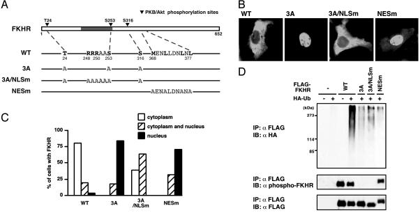
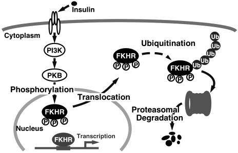
Forkhead transcription factor FKHR (Foxo1) is a key regulator of glucose homeostasis, cell-cycle progression, and apoptosis. It has been shown that FKHR is phosphorylated via insulin or growth factor signaling cascades, resulting in its cytoplasmic retention and the repression of target gene expression. Here, we investigate the fate of FKHR after cells are stimulated by insulin. We show that insulin treatment decreases endogenous FKHR proteins in HepG2 cells, which is inhibited by proteasome inhibitors. FKHR is ubiquitinated in vivo and in vitro, and insulin enhances the ubiquitination in the cells. In addition, the signal to FKHR degradation from insulin is mediated by the phosphatidylinositol 3-kinase pathway, and the mutation of FKHR at the serine or threonine residues phosphorylated by protein kinase B, a downstream target of phosphatidylinositol 3-kinase, inhibits the ubiquitination in vivo and in vitro. Finally, efficient ubiquitination of FKHR requires both phosphorylation and cytoplasmic retention in the cells. These results demonstrate that the insulin-induced phosphorylation of FKHR leads to the multistep negative regulation, not only by the nuclear exclusion but also the ubiquitination-mediated degradation.


Figures

Fig. 1.
Fig. 1.
Insulin induces FKHR turnover through proteasomal degradation. (A and B) HepG2 cells were serum-starved for 12 h and treated with 100 nM insulin (A), with or without a proteasome inhibitor MG132 (B), for the indicated periods. Whole cell extracts were analyzed by immunoblotting. (C) HepG2 cells were serum-starved for 12 h and treated with insulin in the presence or absence of indicated proteasome inhibitors for 12 h. Whole cell extracts were analyzed by immunoblotting.
Fig. 2.
Fig. 2.
FKHR is ubiquitinated in vivo and in vitro. (A) HepG2 cells were transfected with the FLAG-FKHR and HA-ubiquitin expression plasmids. Twenty-four hours after transfection, cells were serum-starved for 12 h and incubated with MG132 with or without insulin or FBS for a further 12 h. Whole cell extracts were subjected to anti-FLAG immunoprecipitation (IP) and followed by anti-HA (Upper) or anti-FLAG (Lower) immunoblotting. (B) HepG2 cells were transfected with the FLAG-FKHR and HA-ubiquitin expression plasmids and treated with MG132 for 10 h before cell lysis. Whole cell extracts were subjected to anti-FLAG immunoprecipitation. The immunoprecipitated materials were eluted from the beads by heating in the presence of 1% SDS and reimmunoprecipitated (re-IP) with the same antibody. All of the immune complexes were analyzed as in A. (C) HepG2 cells transfected with the FLAG-FKHR and HA-ubiquitin were treated with indicated proteasome inhibitors, and ubiquitination was detected as in A.(D) Cell extracts from HEK293T cells transfected with the FLAG-FKHR plasmid were subjected to anti-FLAG immunoprecipitation. The immune complexes were incubated at 30°C for 1 h with or without RRL and either GST or GST-HA-ubiquitin. (Right) The reaction products were eluted by FLAG peptide, again subjected to pull-down by glutathione-Sepharose or to immunoprecipitation by using anti-HA antibody. All of the reaction products were analyzed by immunoblotting with anti-FKHR antibody.
Fig. 3.
Fig. 3.
Phosphorylation via PI3K-PKB pathway is important for FKHR ubiquitination. (A) HepG2 cells were serum-starved for 12 h and preincubated with PI3K inhibitor, LY294002, or wortmannin for 30 min and stimulated with insulin for 6 h. Whole cell extracts were analyzed by immunoblotting using anti-FKHR antibody. (B) FLAG-FKHR and HA-ubiquitin were cotransfected with a dominant negative mutant of PI3K (Xp-Δp85) expression plasmids in HepG2 cells. After treatment with MG132, whole cell extracts were subjected to anti-FLAG immunoprecipitation and followed by immunoblotting. The expression of Xpress-tagged Δp85 in the cell extract was shown by immunoblotting with anti-Xpress antibody. (C) HepG2 cells were transfected with the indicated FKHR mutant and HA-ubiquitin expression plasmids and treated with MG132. Whole cell extracts were subjected to anti-FLAG immunoprecipitation, followed by anti-HA(Upper) or anti-FLAG (Lower) immunoblotting. (D) Cell extracts from HEK293T cells transfected with the FLAG-FKHR WT or 3A plasmid were subjected to anti-FLAG immunoprecipitation. The immune complexes were incubated at 30°C for 1 h in the presence or absence of RRL. The reaction products were analyzed by immunoblotting with anti-FKHR antibody.
Fig. 4.
Fig. 4.
Both phosphorylation and cytoplasmic retention are necessary for efficient FKHR ubiquitination. (A) A schematic representation of FKHR point mutants. The gray box indicates the forkhead DNA binding domain. (B) Localization of FKHR WT or mutants was monitored by transfection of these FKHRs into HepG2 cells in the presence of FBS and by immunolocalization with anti-FLAG antibody. (C) For quantitation, 100 cells per coverslips were counted, and the results are shown as the percentage of cells. (D) HepG2 cells were transfected with the indicated FKHR mutant and HA-ubiquitin expression plasmids and treated with MG132 in the presence of FBS. Whole cell extracts were subjected to anti-FLAG immunoprecipitation and followed by anti-HA (Top), anti-phospho-FKHR (Middle), or anti-FLAG (Bottom) immunoblotting.
Fig. 5.
Fig. 5.
A model for FKHR regulation through insulin-induced and phosphorylation-dependent degradation.

References

    1. Kandel, E. S. & Hay, N. (1999) Exp. Cell Res. 253, 210–229. - PubMed
    1. Kido, Y., Nakae, J. & Accili, D. (2001) J. Clin. Endocrinol. Metab. 86, 972–979. - PubMed
    1. Burgering, B. M. & Kops, G. J. (2002) Trends Biochem. Sci. 27, 352–360. - PubMed
    1. Birkenkamp, K. U. & Coffer, P. J. (2003) Biochem. Soc. Trans. 31, 292–297. - PubMed
    1. Brunet, A., Bonni, A., Zigmond, M. J., Lin, M. Z., Juo, P., Hu, L. S., Anderson, M. J., Arden, K. C., Blenis, J. & Greenberg, M. E. (1999) Cell 96, 857–868. - PubMed

Publication types

MeSH terms