. 2003 Sep 30;100(20):11309-14.
doi: 10.1073/pnas.1834451100. Epub 2003 Sep 17.

Electron microscopic evidence for nucleation and growth of 3D acetylcholine receptor microcrystals in structured lipid-detergent matrices

Affiliations

Electron microscopic evidence for nucleation and growth of 3D acetylcholine receptor microcrystals in structured lipid-detergent matrices

Yoav Paas et al. Proc Natl Acad Sci U S A. .

Abstract

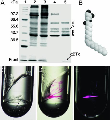
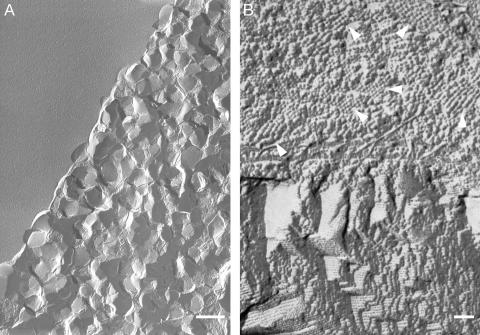
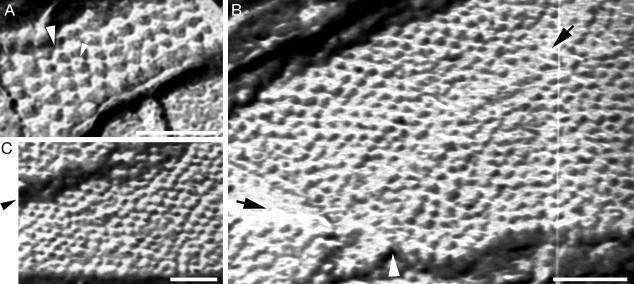
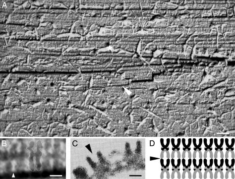
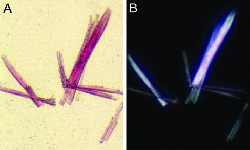
Nicotinic acetylcholine receptors (AChRs) belong to a superfamily of oligomeric proteins that transduce electric signals across the cell membrane on binding of neurotransmitters. These receptors harbor a large extracellular ligand-binding domain directly linked to an ion-conducting channel-forming domain that spans the cell membrane 20 times and considerably extends into the cytoplasm. Thus far, none of these receptor channels has been crystallized in three dimensions. The crystallization of the AChR from Torpedo marmorata electric organs is challenged here in lipidic-detergent matrices. Detergent-soluble AChR complexed with alpha-bungarotoxin (alphaBTx), a polypeptidic competitive antagonist, was purified. The AChR-alphaBTx complex was reconstituted in a lipidic matrix composed of monoolein bilayers that are structured in three dimensions. The alphaBTx was conjugated to a photo-stable fluorophore, enabling us to monitor the physical behavior of the receptor-toxin complex in the lipidic matrix under light stereomicroscope, and to freeze fracture regions containing the receptor-toxin complex for visualization under a transmission electron microscope. Conditions were established for forming 2D receptor-toxin lattices that are stacked in the third dimension. 3D AChR nanocrystals were thereby grown inside the highly viscous lipidic 3D matrix. Slow emulsification of the lipidic matrix converted these nanocrystals into 3D elongated thin crystal plates of micrometer size. The latter are stable in detergent-containing aqueous solutions and can currently be used for seeding and epitaxial growth, en route to crystals of appropriate dimensions for x-ray diffraction studies.


Figures

Fig. 1.
Fig. 1.
Incorporation of AChR–αBTx complexes into a lipidic 3D matrix. (A) Reducing SDS-polyacrylamide gel (9%) showing the purification steps: lane 1, molecular mass markers; lane 2, receptor-rich membranes; lane 3, pH 11-treated receptor-enriched membranes; lane 4, CHAPS-solubilized receptor; lane 5, purified fraction. αBTx (8.5 kDa) that migrates at the front and the receptor subunits are indicated. Shown is a gel stained by Coomassie brilliant blue. (B) Space-filling model of a MO molecule. Dark and light spheres correspond to oxygen and carbon atoms, respectively. (C) MO mixed with the protein-carrier buffer at MO:buffer ratio of 6:4 (wt/wt). The inner diameter of the glass tube is 2.5 mm. (D) Example of purified AChR–αBTx complexes concentrated inside the lipidic phase; the red color corresponds to tetramethylrhodamine that is covalently attached to the toxin. Shown is a picture taken under regular light. (E) The same sample as shown in D but under polarized light. Note that only regions that contain receptor–toxin complexes become birefringent. Also, there are regions that are red but not birefringent; their portion may vary between ≈5% and 40% of the total incorporated AChR–αBTx.
Fig. 2.
Fig. 2.
Transmission electron micrographs of regions containing AChR–αBTx complexes. (A) Freeze fracture of red-labeled birefringent regions such as those shown in Fig. 1 D and E reveals many nanocrystals. Note that the upper left corner corresponds to a flat lipidic surface devoid of nanocrystals. (Scale bar = 0.5μm.) (B) Top view of a freeze-fracture plane showing segregation between particle-containing (upper part) and particle-devoid (lower part) regions. The arrows indicate six of numerous small regions consisting of spherical particles that are densely packed as highly ordered aggregates reminiscent of nuclei and tiny nanocrystals. The terrace-like architecture depicted in the lower part corresponds to ≈4-nm-width layers stacked on top of each other. (Scale bar = 50 nm.) The white/grayish areas are shadowed by platinum.
Fig. 3.
Fig. 3.
AChR–αBTx complexes are organized in 2D lattices. (A) Top view of a nanometer-sized crystal reflecting nucleation of receptor–toxin complexes inside the MO cubic phase. The small arrow indicates a receptor molecule that appears like a doughnut displaying five subunits surrounding a black pit. The diameter of this receptor is ≈9 nm. The large arrow indicates a receptor molecule that collapsed and coalesced with the neighboring right-handed receptor. (B) Top view of a region showing hundreds of spherical particles organized side by side in linear rows. These particles correspond to AChR molecules, as discussed in the text. The black arrow in the upper right corner points to a defect propagating along the nanocrystal. The black and white arrows at the bottom left corner pointto a contact area between two nanocrystals. Regular facets can be seen at the upper and lower edges. (C) A wide crack along a nanocrystal is pointed by the arrow. A regular facet can be visualized at the lower edge. In all panels, the scale bars correspond to 50 nm, and the white/grayish areas are shadowed by platinum.
Fig. 4.
Fig. 4.
AChR–αBTx complexes are organized in layers stacked in the third dimension. (A) Freeze-fracture plane passing through a mid-longitudinal axis of the receptor molecules exposes a multilayer organization. The grayish ruptures (one is indicated by a white asterisk) correspond to pieces that were torn apart from the fracture plane. The region indicated by the white arrow is magnified in B. Note that many receptor molecules collapsed, probably because of the fracturing through hard proteinaceous material. The receptor molecules are darkened, and the pits are white/grayish. (Scale bar = 50 nm.) (B) Magnification of six “U-shaped” constituents of the region indicated in A by a white arrow. These receptor molecules are arranged side by side and are tightly packed tail to tail. For convenience, a white line delineates the boundaries of the middle receptor dimer. The white arrow indicates the pit of a receptor molecule as can be compared with the pit shown in C. (Scale bar = 50 Å.) (C) Side view of AChR molecules as seen in a portion of a tubular 2D crystal previously obtained from pH 11-treated membranes. [Reproduced with permission from ref. (Copyright 1988, Nature Publishing Group, www.nature.com).] The arrow points to the receptor's pit. (Scale bar = 50 Å.) (D) Scheme illustrating the packing pattern of the receptor molecules as concluded from A and B. The arrow indicates an initiation point for a fracture that would provide a top view of the lattice, as observed in many fracture planes (e.g., Fig. 3).
Fig. 5.
Fig. 5.
Emulsification of the lipidic phase results in growth of micrometersized AChR–αBTx crystals. (A) A few thin crystal plates that grew on slow liquefaction of MO cubic phase that harbored AChR–αBTx nanocrystals. Shown is a picture taken under a regular light. The dimensions of the largest crystal are: 300 μm × 30 μm ×≈15 μm. (B) The same crystals as shown in A but under polarized light.

Similar articles

Cited by

References

    1. Rosenbusch, J. P. (2001) J. Struct. Biol. 136, 144–157. - PubMed
    1. Corringer, P. J., Le Novere, N. & Changeux, J. P. (2000) Annu. Rev. Pharmacol. Toxicol. 40, 431–458. - PubMed
    1. Karlin, A. (2002) Nat. Rev. Neurosci. 3, 102–114. - PubMed
    1. Engel, A. G., Ohno, K. & Sine, S. M. (2003) Nat. Rev. Neurosci. 4, 339–352. - PubMed
    1. Kistler, J. & Stroud, R. M. (1981) Proc. Natl. Acad. Sci. USA 78, 3678–3682. - PMC - PubMed

Publication types