Review
. 2003 Sep 30;100 Suppl 1(Suppl 1):11917-23.
doi: 10.1073/pnas.1834138100. Epub 2003 Sep 17.

One strategy for cell and gene therapy: harnessing the power of adult stem cells to repair tissues

Affiliations
Review

One strategy for cell and gene therapy: harnessing the power of adult stem cells to repair tissues

Darwin J Prockop et al. Proc Natl Acad Sci U S A. .

Abstract

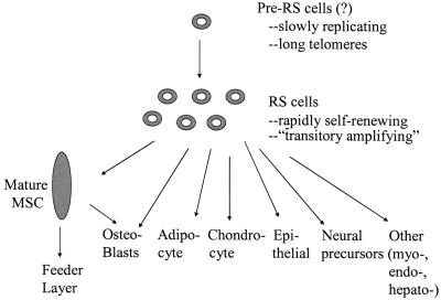
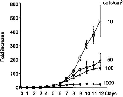
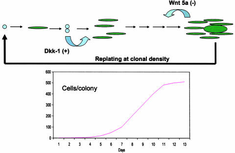
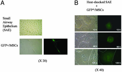
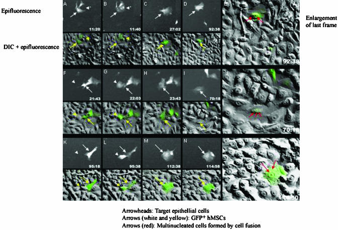
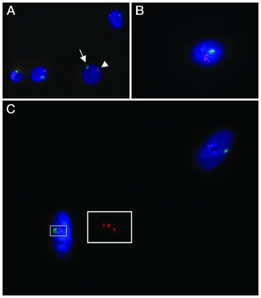
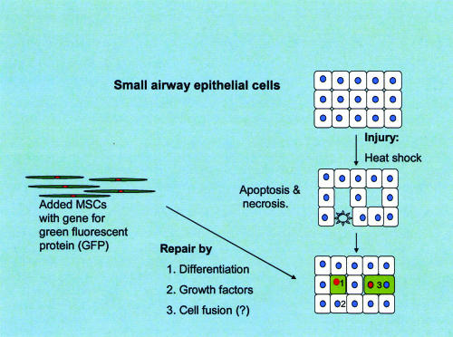
Most recent evidence suggests that the process of tissue repair is driven by stem-like cells that reside in multiple tissues but are replenished by precursor cells from bone marrow. Among the candidates for the reparative cells are the adult stem cells from bone marrow referred to as either mesenchymal stem cells or marrow stromal cells (MSCs). We recently found that after MSCs were replated at very low densities to generate single-cell-derived colonies, they did not exit a prolonged lag period until they synthesized and secreted considerable quantities of Dickkopf-1, an inhibitor of the canonical Wnt signaling pathway. We also found that when the cells were cocultured with heat-shocked pulmonary epithelial cells, they differentiated into epithelial cells. Most of the MSCs differentiated without evidence of cell fusion but up to one-quarter underwent cell fusion with the epithelial cells. A few also underwent nuclear fusion. The results are consistent with the interesting possibility that MSCs and similar cells repair tissue injury by three different mechanisms: creation of a milieu that enhances regeneration of endogenous cells, transdifferentiation, and perhaps cell fusion.


Figures

Fig. 1.
Fig. 1.
Schematic summarizing relationship between subpopulations of MSCs and their differentiation to specific cell phenotypes.
Fig. 2.
Fig. 2.
Lag period and rapid expansion of early-passage human MSCs plated at low densities. [Reproduced with permission from ref. (Copyright 2002, AlphaMed Press).]
Fig. 3.
Fig. 3.
Scheme summarizing transitions of colonies of MSCs from lag period to log phase and stationary phase of growth. As indicated, Dkk-1 is synthesized and secreted as late log/early log phase and stimulates expansion of cells. Wnt 5a in expressed at the end of log phase and in stationary phase. As the cells expand in the colonies, spindle-shaped RS cells give rise to larger, more mature cells. For four or five passages of preparation of MSCs the sequence is repeated after the cells are lifted and replated at low density.
Fig. 4.
Fig. 4.
Cocultures of GFP+ MSCs and heat-shocked SAECs. (A) The SAECs form a continuous monolayer of epithelial cells that are broad and thin with a raised perinuclear region. In the serum-free medium used to culture SAECs, GFP+ MSCs become thin and elongated (epifluorescence frame, Lower Right). (B) Differential interference (Left) and epifluorescence (Right) of cocultures. After heat shock, the monolayer of SAECs is disrupted as some of the cells undergo necrosis and apoptosis. Over 12-120 h, some GFP+ MSCs enter the monolayer and become broad, flat cells that reform the monolayer. (Outline of GFP+ cell in Bottom is enhanced.) [Reproduced with permission from ref. (Copyright 2003, National Academy of Sciences).]
Fig. 5.
Fig. 5.
Time-lapse photomicroscopy of cocultures of GFP-labeled human MSCs added to heat-shocked SAECs demonstrating cell fusion. Epifluorescence and differential interference (DIC) photomicrographs were taken every 20 min for >114 h, and selective frames for three sequences are shown. In alternate rows, the epifluorescence and DIC frames are superimposed. [Reproduced with permission from ref. (Copyright 2003, National Academy of Sciences).]
Fig. 6.
Fig. 6.
Evidence for nuclear fusion in cocultures of GFP-labeled male MSCs and heat-shocked female SAECs. After coculture, fluorescence-activated cell sorting was used to isolate cells positive both for GFP and CD24, a surface epitope expressed on the epithelial cells but not on MSCs. The isolated cells were then assayed by in situ hybridization for the X and Y chromosome. (A) Control male cell (arrows) containing one X and one Y chromosome. (B) A rare cell with a single nucleus containing one Y chromosome and five X chromosomes. (C) A highlighted cell with a single nucleus containing one Y and three X chromosomes. (Inset) Enlargement to demonstrate the three X chromosomes. The second nucleus has one X and one Y chromosome, an observation consistent with differentiation without cell fusion of a male MSC.
Fig. 7.
Fig. 7.
Schematic showing that in the coculture system some MSCs differentiated directly into SAECs and others fused. In addition, the MSCs probably synthesize and secrete growth factors that enhance regeneration of epithelial and other cells.

References

    1. Tanaka, E. M. (2003) Cell 113, 559-562. - PubMed
    1. Cohnheim, J. (1867) Pathol. Anat. Physiol. Klin. Med. 40, 1-79.
    1. Ross, R., Everett, N. B. & Tyler, R. (1970) J. Cell Biol. 44, 645-654. - PMC - PubMed
    1. Prockop, D. J. (1997) Science 276, 71-74. - PubMed
    1. Doherty, M. J. & Canfield, A. E. (1999) Crit. Rev. Eukaryotic Gene Expression 9, 1-17. - PubMed

Publication types