. 1992 Mar 5;267(7):4930-8.

Biochemical properties of chimeric skeletal and smooth muscle myosin light chain kinases

Affiliations

Biochemical properties of chimeric skeletal and smooth muscle myosin light chain kinases

S A Leachman et al. J Biol Chem. .

Abstract

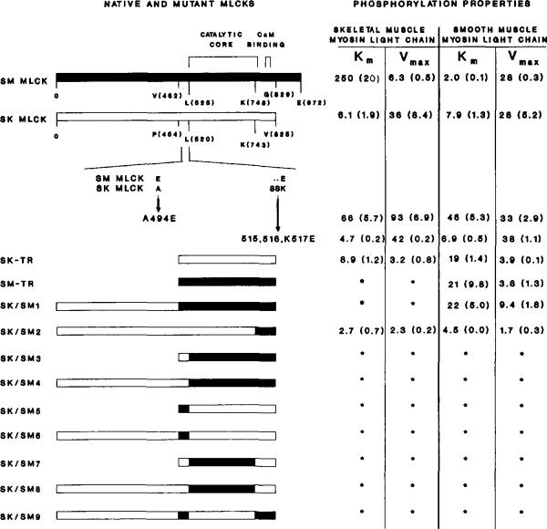
The molecular and biochemical properties of myosin light chain kinases from chicken skeletal and smooth muscle were investigated by recombinant DNA techniques. Deletion of the amino-terminal region of either the smooth or skeletal muscle myosin light chain kinase resulted in a decrease in Vmax with no significant change in Km values for light chain substrates. Skeletal/smooth muscle chimeric kinases were inactive when a 65-residue region amino-terminal of the catalytic core was exchanged between the two forms. Changing alanine 494 to glutamic acid within this region in the chicken skeletal muscle myosin light chain kinase increased the Km values for light chains 10-fold. These results are consistent with the hypothesis that the region amino-terminal of the catalytic core in myosin light chain kinases is involved in light chain recognition. A skeletal muscle kinase which contained the smooth muscle calmodulin binding domain remained regulated by Ca2+/calmodulin. Thus, the calmodulin binding domains of smooth and skeletal muscle myosin light chain kinases share structural elements necessary for regulation.


Figures

Fig. 1
Fig. 1
Panel A, partial restriction map of the cDNA encoding chicken skeletal muscle MLCK. Restriction endonuclease sites that were used in the construction of the cDNA, truncation mutants, or chimeric MLCKs are indicated at the following positions in the clone: SmaI (141), NcoI (185), ApaI (1546), BglII (2039), StyI (2411), and PstI (2480). EcoRI sites originate from linkers used to prepare the libraries; there are no natural EcoRI sites in the cDNA. The synthetic oligonucleotide primer and its relative position are indicated by the solid black bar at the 3′ end of the 2.1-kb partial clone. Directional arrows indicate individual sequencing reactions. Panel B, construction of truncation and chimeric cDNAs. Oligonucleotide sequences and their relative positions are indicated on the linear schematic of the nucleotide sequence. Numbers correspond to the nucleotides of the chicken skeletal (Fig. 2) or smooth (Olson et al., 1990) muscle cDNAs on the respective cDNAs.
Fig. 2
Fig. 2. Nucleotide and deduced amino acid sequence of the cDNA encoding chicken skeletal muscle MLCK
The nucleotide sequence and the deduced amino acid sequence of the coding region of the chicken skeletal muscle MLCK are shown in capital letters. The 5′-untranslated region has been translated in lowercase letters. The initiating M is in boldface type. Aspartyl-prolyl bonds are indicated with asterisks as potential sites for cleavage by 70% formic acid. The overlined residues indicate the region amino-terminal of the catalytic core which is homologous to other MLCKs (residues 479–511). The catalytic core of the enzyme is bracketed (residues 512–772), and the calmodulin binding domain is underlined (residues 795–811).
Fig. 3
Fig. 3. Comparison of the primary structures of MLCKs from different tissues and species
Panel A, a linear schematic shows full-length MLCKs (amino-terminal to carboxyl-terminal) from rat skeletal muscle (RATSKMLCK; Roush et al., 1988), rabbit skeletal muscle (RABSKMLCK; Takio et al., 1986), chicken skeletal muscle, chicken smooth muscle (CHKSMMLCK; Olson et al., 1990), and chicken fibroblasts (CHKNMMLCK; Shoemaker et al., 1990). The solid block indicates the position of the conserved catalytic cores. The shaded region amino-terminal of the catalytic cores of the skeletal muscle enzymes indicates a region of extended homology among these MLCKs. The shqded region carboxyl-terminal of the catalytic core indicates the position of the calmodulin binding domains. Panel B, amino acid sequence alignment of the same MLCKs is shown beginning with the region that is conserved amino-terminal of the catalytic core. A consensus sequence is shown above the sequence alignment. Within the consensus sequence, identical residues are indicated by capital letters and conservative substitutions by solid ovals. Conservation is based on the following groupings of structurally similar amino acids: basic polar R groups (K, R, and H); acidic and uncharged polar R groups (D, E, N, and Q); nonpolar chain R groups (M, L, I, and V); aromatic or ring-containing R groups (F, Y, W, and H); and small R groups with near neutral polarity (A, C, G, S, T, and P). Subdomains I-XI of the catalytic core (Hanks et al., 1988) and the calmodulin binding domain are bracketed above the consensus sequence. Residues that are conserved among all protein kinases are underlined in the consensus sequence. The GXGXXG consensus sequence found among nucleotide-binding proteins is within subdomain I. Subdomain II contains an invariant lysine among all protein kinases. Residues that are identical among all the skeletal muscle enzymes and different from the smooth and non-muscle enzyme are shaded, including the carboxyl-terminal extension of the smooth and non-muscle enzymes which are uniformly absent in the skeletal muscle enzymes.
Fig. 4
Fig. 4. Western immunoblot and calmodulin overlay analysis of chicken skeletal and smooth muscle MLCKs and mutant enzymes
Purified MLCKs and COS cell extracts containing recombinant proteins were separated by 7.5% SDS-PAGE and detected with monoclonal antibodies or biotinylated calmodulin as described under “Materials and Methods.” Panel A, Western immunoblot with monoclonal antibody raised to chicken skeletal muscle MLCK. Lane 1, mock transfected COS cell lysate; lane 2, tissue-purified chickn skeletal muscle MLCK (10 ng); lanes 3–11, COS cell lysates transfected with 3, wild-type chicken skeletal muscle MLCK, 4, SK/SM1; 5, SK/SM4; 6, SK/SM6; 7, SK/SM8; 8, SK/SM2; 9, SK/SM9; 10, A494E 11, Δ515–516,K517E. Panel B, biotinylated calmodulin overlay. Lane 1, mock transfected COS cell lysate; lane 2, tissue-purified chicken skeletal myosin light chain kinase; lanes 3–6, COS cell lysates with 3, wild-type chicken skeletal muscle myosin light chain kinase; 4, SK-TR mutant; 5, SK/SM1 mutant; 6, SM-TR mutant.
Fig. 5
Fig. 5. Linear schematic representation of the primary structures and kinetic properties of chicken skeletal and smooth muscle native, truncated, and chimeric MLCKs
Regions representing skeletal muscle myosin light chain kinase (SK MLCK) are outlined, and regions from smooth muscle kinase (SM MLCK) are darkly shaded. Km (μM) and Vmax (μmol of 32P incorporated per min/mg) values for skeletal and smooth muscle myosin light chains are shown to the right of the native or mutant MLCKs. The numbers represent the means with the S.E. in parentheses. The positions of the catalytic core and calmodulin binding domain are indicated above the linear schematic. The smooth muscle MLCK has 972 residues: residues 519–774 form the catalytic core and residues 797–813 form the calmodulin binding domain. The native skeletal muscle enzyme is composed of 825 amino acids with the catalytic core and calmodulin binding domain spanning residues 512–772 and 795–811, respectively. An amino acid sequence alignment below the native skeletal muscle MLCK illustrates the site-specific mutants A494E and Δ515–516,K517E. The compositions of the truncation mutants and chimeric enzymes are as follows (numbers in parentheses are amino acid numbers): SK-TR = SK (453–825); SM–TR = SM (462–829); SK/SM1 = SK (1–454) and SM (462–829); SK/SM2 = SK (1–742) and SM (748–829); SK/SM3/B> = SK (453–520) and SM (525–829); SK/SM4 = SK (1–520) and SM (526–829); SK/SM5 = SK (453–454), SM (462–525), and SK (521–825); SK/SM6 = SK (1–454), SM (462–525), and SK (521–825); SK/SM7 = SK (453–520), SM (526–748), and SK (744–825); SK/SM8 = SK (1–520), SM (526–748), and SK (744–825); SM (SK/SM9) = SK (1–454), SM (462–525), SK (521–744), and SM (750–829).

Similar articles

Cited by

References

    1. Adelstein RS, Klee CB. J. Biol. Chem. 1981;256:7501–7509. - PubMed
    1. Andersson S, Davis DL, Dahlbäch H, Jörnvall H, Russell DW. J. Biol. Chem. 1989;264:8222–8229. - PubMed
    1. Bagchi IC, Kemp BE, Means AR. J. Biol. Chem. 1989;264:15843–15849. - PubMed
    1. Billingsley ML, Pennypacker KR, Hoover CG, Brigati DJ, Kincaid RL. Proc. Natl. Acad. Sci. U. S. A. 1985;82:7585–7589. - PMC - PubMed
    1. Blumenthal DK, Stull JT. Biochemistry. 1982;21:2386–2391. - PubMed

Publication types

MeSH terms

LinkOut - more resources