

doi: 10.1016/0014-4800(62)90020-5.
Histopathologic alterations associated with the transplanted homologous dog liver
- PMID: 13873233
- PMCID: PMC3005267
- DOI: 10.1016/0014-4800(62)90020-5
Item in Clipboard
Histopathologic alterations associated with the transplanted homologous dog liver
Exp Mol Pathol.
1962 Jun.
No abstract available
Figures
Liver from dog surviving 10 hours. Note portal venous and lymphatic engorgement characteristic of outflow block, × 43.
Liver from dog surviving 10 hours illustrating Kupfer cell activation, × 263.
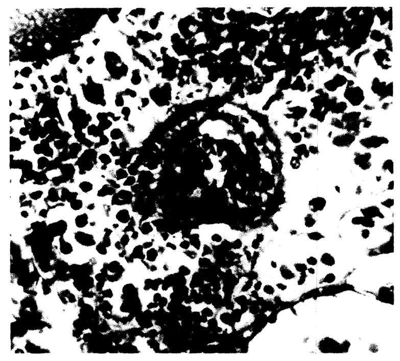
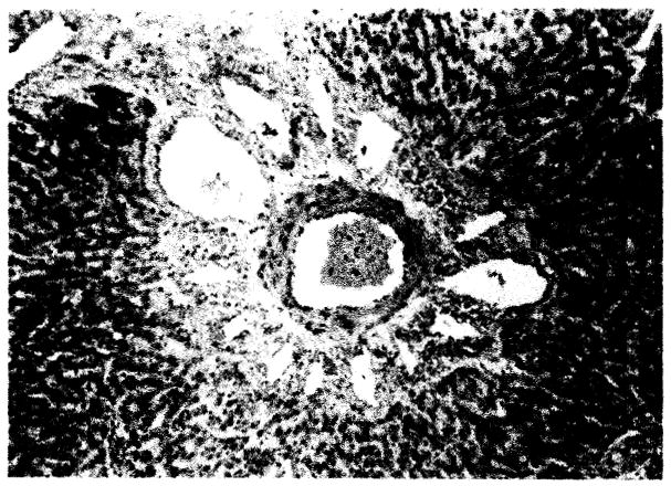
Arteriole of liver showing endothelial and perithelial proliferation with focal hyaline degeneration, × 263.

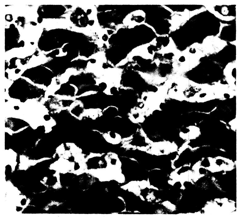
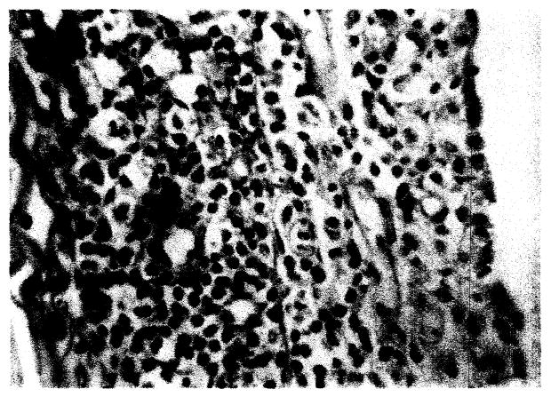
Liver: 10 day survival, × 263. Periportal and parenchymal infiltrate is primarily plasmacytic.
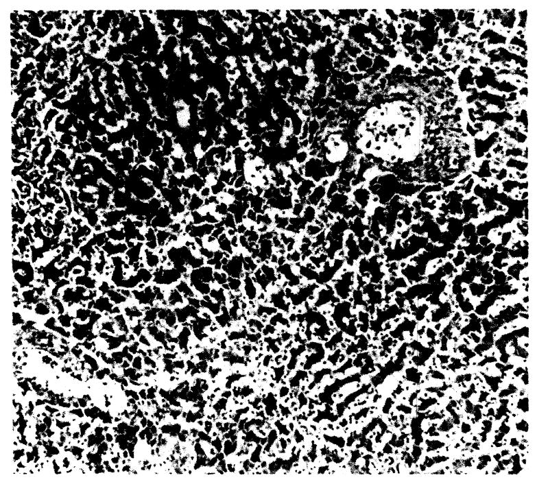
General hepatic architecture: (a) 9 day survival, × 42; (b) another dog, 8 day survival, × 42.
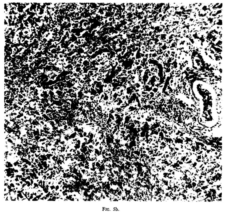
General hepatic architecture: (a) 9 day survival, × 42; (b) another dog, 8 day survival, × 42.
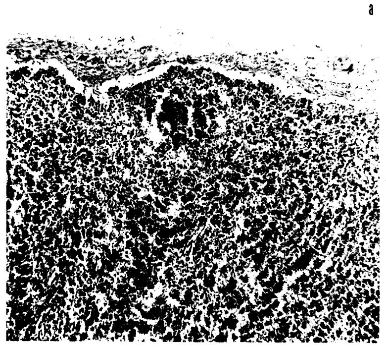
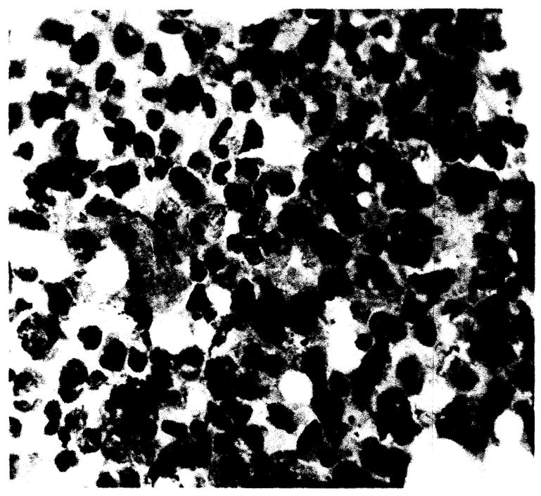
Lymph node from dog surviving 20 days: (a) cortical depletion, × 82; (b) cortical cell type primarily mononuclear and plasmacytic, × 232.
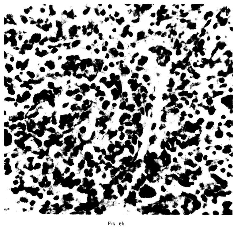
Lymph node from dog surviving 20 days: (a) cortical depletion, × 82; (b) cortical cell type primarily mononuclear and plasmacytic, × 232.
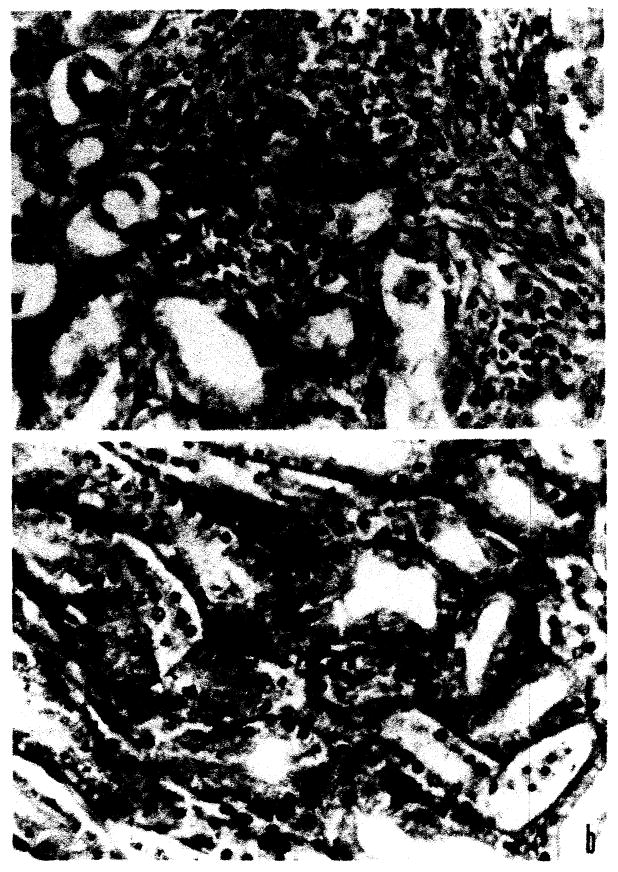
Kidney: (a) plasmacytic aggregation, × 97; (b) tubular basement membrane activation, × 97.
Perirenal plasmacytic and mononuclear infiltration, × 97.
Lung. Focal alveolar wall proliferation, mononuclear cell infiltrate and giant cell formation, × 225.
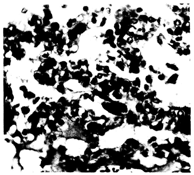
Bone marrow. Note increased plasmacytes, × 392.
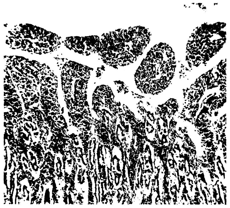
Small intestine. Mucosal slough and mononuclear infiltration, × 42.
References
-
- Bjornboe M, Gormsen H. Experimental studies on the role of plasma cells as antibody producers. Acta Pathol Microbiol Scand. 1943;20:649–692. - PubMed
-
- Dempster WJ. Kidney homotransplantation. Brit J Surg. 1953;40:447–465. - PubMed
-
- Kojima M. Morphologic changes accompanying RES stimulation. Ann NY Acad Sci. 1960;88(1):196–202. - PubMed
MeSH terms
Grants and funding
LinkOut - more resources
Full Text Sources
