

doi: 10.1016/0002-9610(62)90491-9.
Homotransplantation of multiple visceral organs
- PMID: 13916395
- PMCID: PMC2998393
- DOI: 10.1016/0002-9610(62)90491-9
Item in Clipboard
Homotransplantation of multiple visceral organs
Am J Surg.
1962 Feb.
No abstract available
Figures
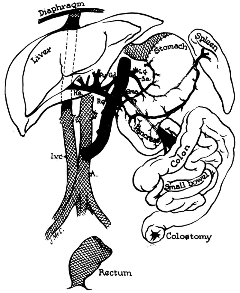
Schematic view of the transplanted tissues and their anatomic relation to the host. The grafted tissues are not shaded.
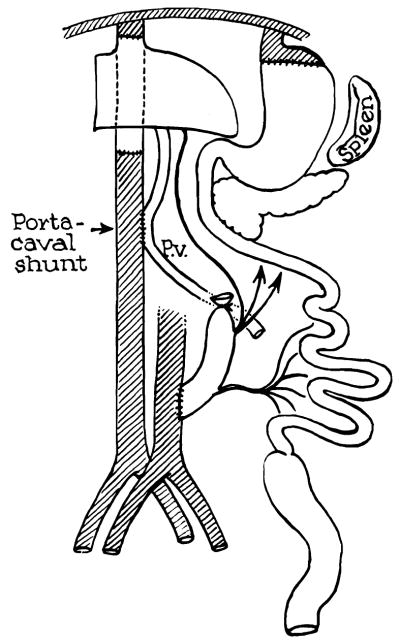
Addition of portacaval shunt to operation depicted in Figure 1.
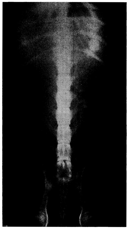
Abdominal roentgenogram of Dog No. 18 on the sixth postoperative day. Dog had been on oral intake for four days.
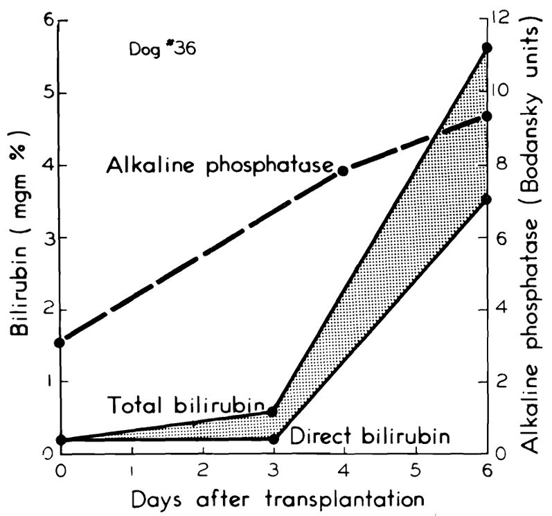
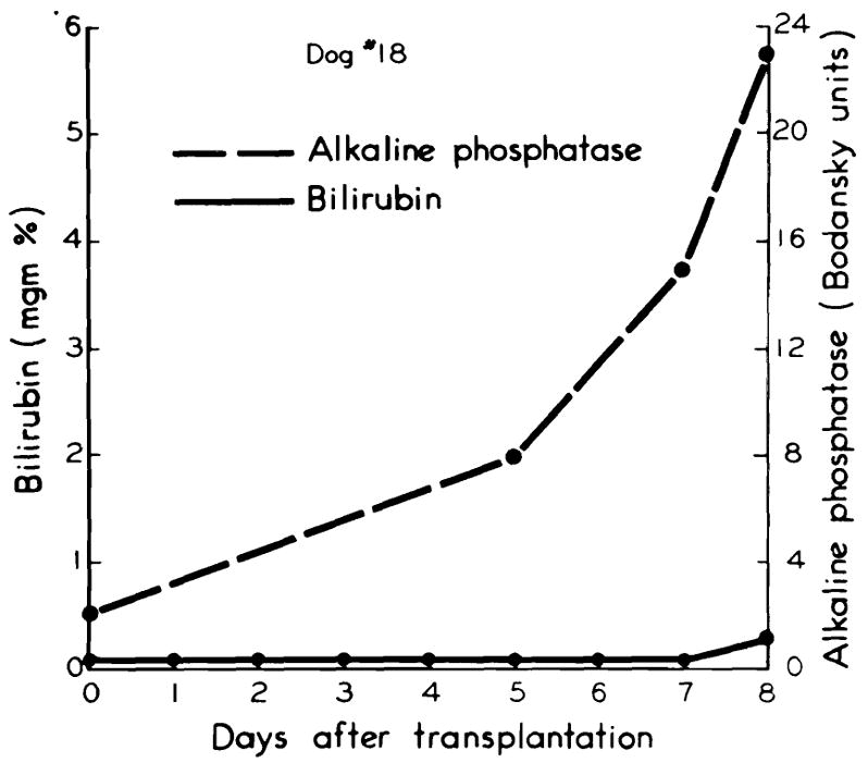
Development of chemical jaundice with rise in alkaline phosphatase, seen in three of the five long-surviving dogs.
Absence of jaundice in two of the five long-surviving dogs. Note rise in alkaline phosphatase.
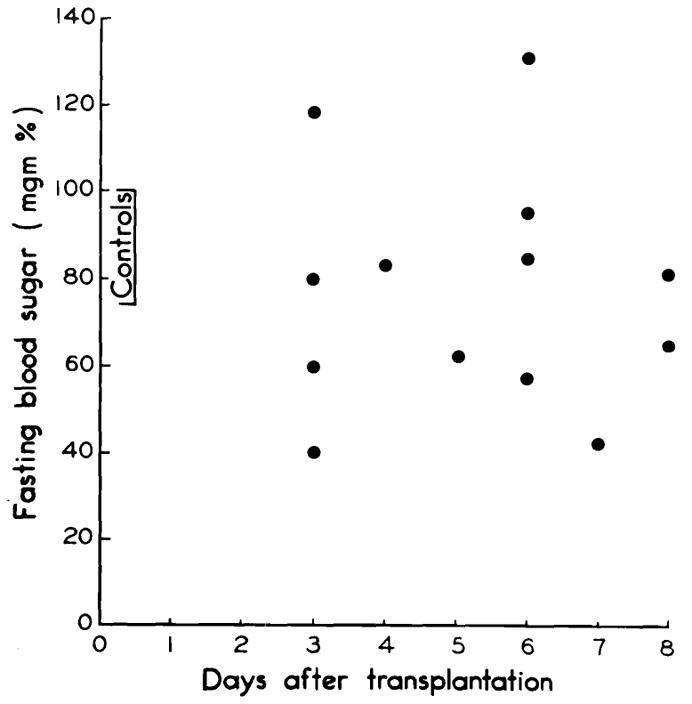
Fasting blood sugars in the five long-surviving dogs. Note usual absence of pronounced hypoglycemia.
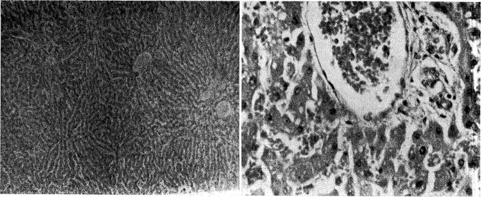
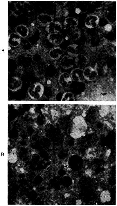
Liver after nine days, from Dog No. 18, in which jaundice did not develop. A, magnification × 65; B, magnification × 350.
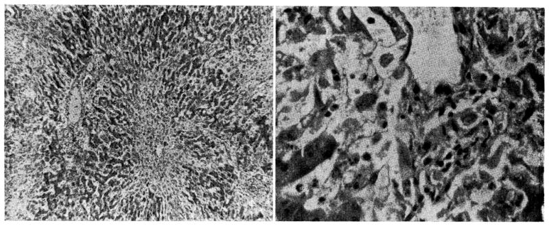
Liver after seven days, from Dog No. 4, in which jaundice developed. A, magnification × 65; B, magnification × 350.
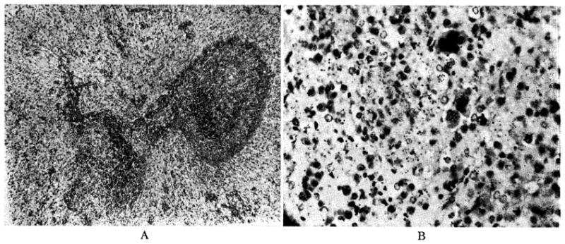
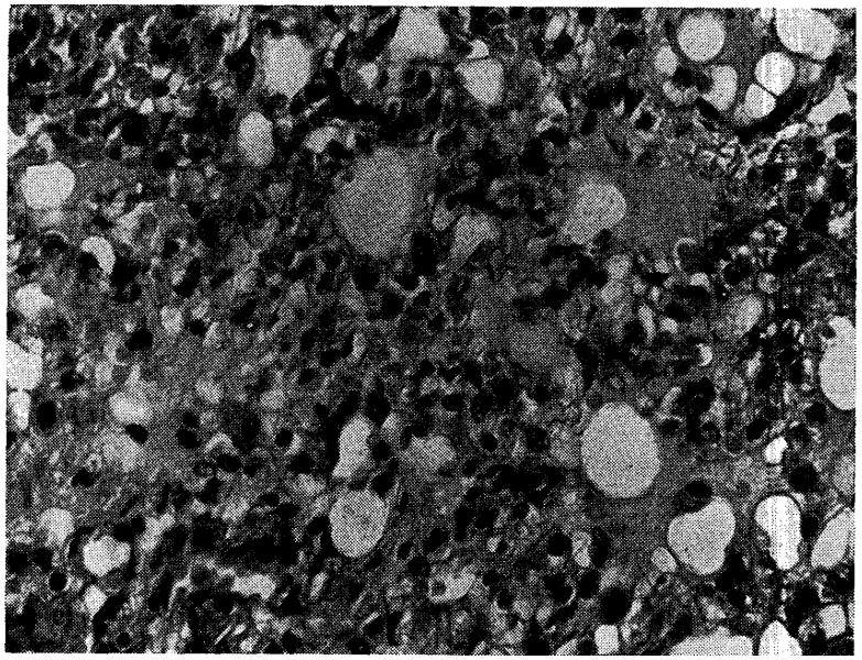
Donor spleen, after nine days, from Dog No. 19. A, magnification × 65; B, magnification × 350.
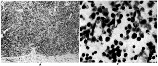
Donor lymph node from mesentery of graft in Dog No. 26. Animal lived five and a half days. A, magnification × 30; B, magnification × 600.
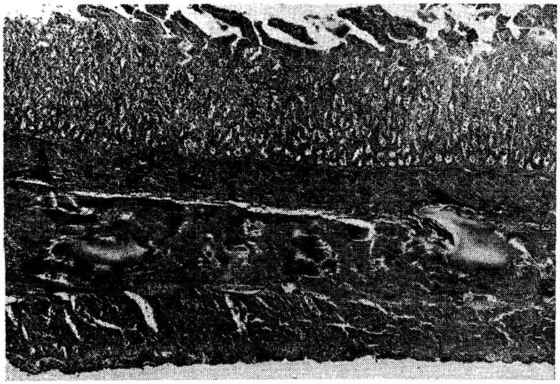
Small intestine of Dog No. 4, seven days after transplantation (magnification × 18). Note congestion, edema and superficial slough.
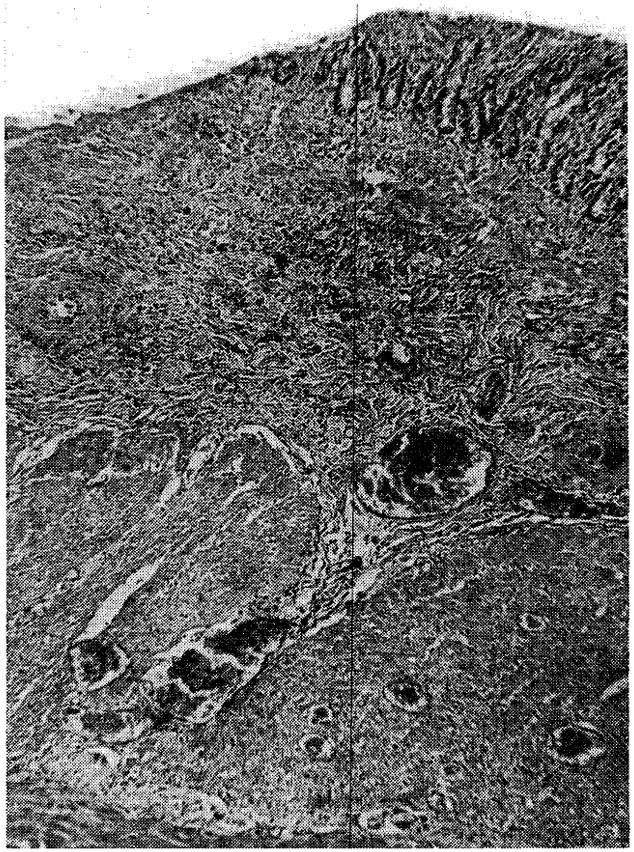
Duodenal ulcer in Dog No. 18, after nine days (magnification × 25).
A, bone marrow from normal dog showing active granulopoiesis and erythropoiesis (magnification × 900). B, marrow from Dog No. 26, showing a cellular specimen with extensive replacement of normal myeloid elements by a relative and absolute increase in lymphocytes, reticulum cells and plasma cells (magnification × 900).
Lung from Dog No. 18, nine days after visceral transplantation (magnification × 350). Note pulmonary edema and proliferative thickening of alveolar septa.
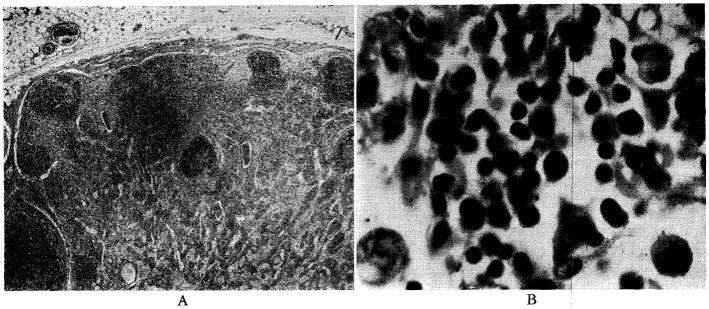
Recipient lymph node from mediastinum of Dog No. 36 after six days. A, magnification × 30; B, magnification × 600.
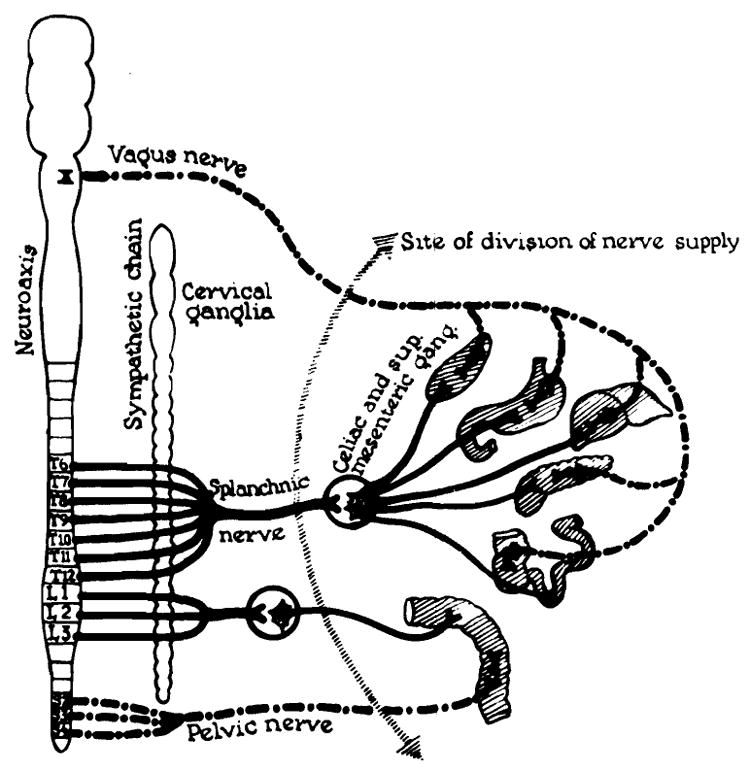
State of denervation of multiple organ graft.
References
-
- Billingham RE. Reactions of grafts against their hosts. Science. 1959;130:947. - PubMed
-
- Goodrich EO, Welch HF, Nelson JA, Beecher TS, Welch CS. Homotransplantation of the canine liver. Surgery. 1956;39:244. - PubMed
-
- Lillehei CW, Wangensteen OH. Effect of celiac ganglionectomy upon experimental peptic ulcer formation. Proc Soc Exper Biol & Med. 1948;68:369. - PubMed
MeSH terms
Grants and funding
LinkOut - more resources
Full Text Sources
