

PHYSIOLOGIC REQUIREMENTS FOR AUXILIARY LIVER HOMOTRANSPLANTATION
- PMID: 14314095
- PMCID: PMC2676919
Item in Clipboard
PHYSIOLOGIC REQUIREMENTS FOR AUXILIARY LIVER HOMOTRANSPLANTATION
Surg Gynecol Obstet.
1965 Jul.
No abstract available
Figures
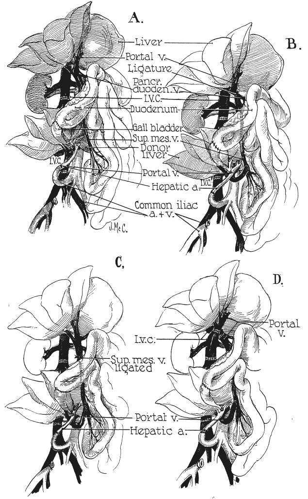
Experimental protocols for auxiliary hepatic homotransplantation. A, Previously reported modification of Welch-Goodrich hepatic homotransplantation. Homograft undergoes rapid atrophy and diminution in size. Portal blood flow is from the systemic venous system. B, Preparation of group 1 dogs in present study. Non-hepatic splanchnic flow is diverted through the homograft. With this preparation, the homograft retains its size and the animal’s own liver undergoes shrinkage. It is usually more convenient to bring the hepatic artery behind, rather than in front of, the portal vein as depicted. C, Superior mesenteric vein is ligated below the splenic vein, partitioning nonhepatic splanchnic flow between the autologous liver and the homograft. Homograft shrinkage occurs but more variably (group 2). D, Auxiliary homotransplantation in group 3. The host liver is vascularized as with the portacaval transposition of Child.
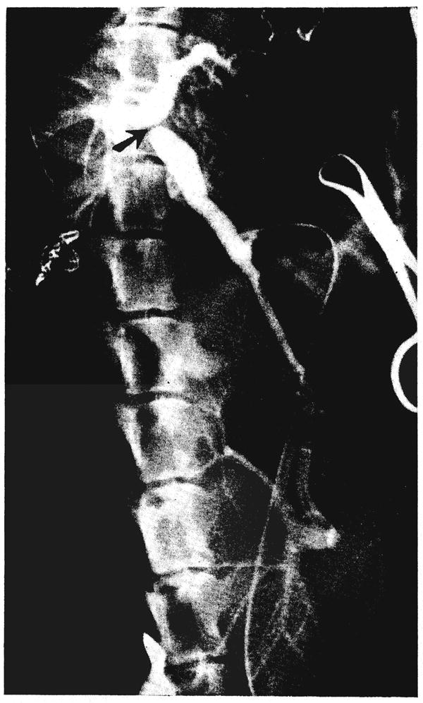
Operative venous angiogram of dog 5 showing retrograde passage of nonhepatic splanchnic flow through the homotransplanted liver (lower). The ligature around the portal vein is evident (arrow). The dog’s own portal system has also filled with dye from collaterals. Note the large size of the homograft compared to the small dimensions of the dog’s own liver.
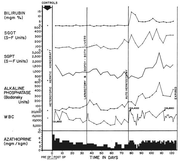
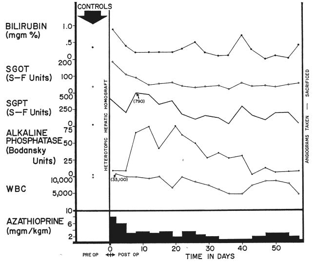
Clinical course of dog 5 of group 1 series. Note the abrupt bilirubinemia which followed removal of the dog’s own liver, autohepatectomy. After autologous hepatectomy, the dog lived for 49 days with sole dependence upon the homograft, ultimately dying as the result of a wound dehiscence and evisceration which followed repeat biopsy.
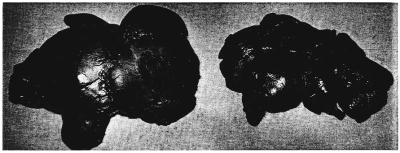
Auxiliary liver homograft, left, compared to the animal’s own liver, right, in dog 8 of group 1 series. The homograft was obtained at autopsy, and the animal’s own liver was removed 8 days earlier. The weight of the homograft was more than twice that of the autologous hepatic tissue.
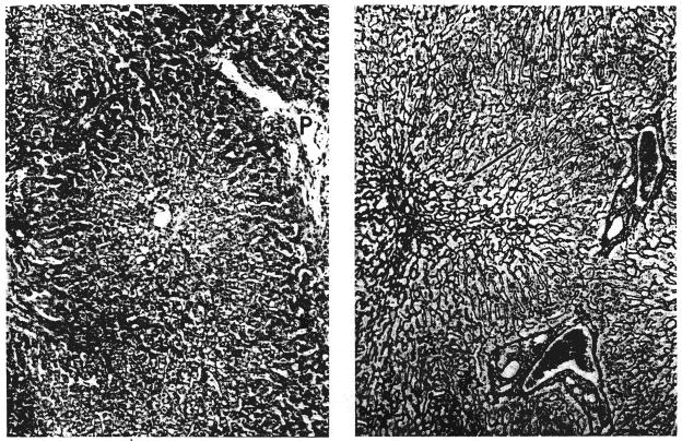
Biopsies of their own livers from 2 dogs in group 1: A, left, 35 days after operation (dog 5). There is centrizonal necrosis of the liver cells; P, portal tract. Hematoxylin and eosin, × 65. B, right, 51 days after operation (dog 10). The centrilobular reticulin has collapsed (arrow). Reticulin, × 90.
Biopsies of hepatic homografts from 2 dogs of group 1: A, left, 35 days after transplantation (dog 5). The portal tracts contain many infiltrating cells. The wall of the central hepatic vein is thickened and lightly infiltrated with cells. Hematoxylin and eosin, × 65. B, right, 61 days after transplantation (dog 8). There is a dense accumulation of reticulin and collagen fibers around and within the wall of the central hepatic vein; P, portal tract. Reticulin, × 65.
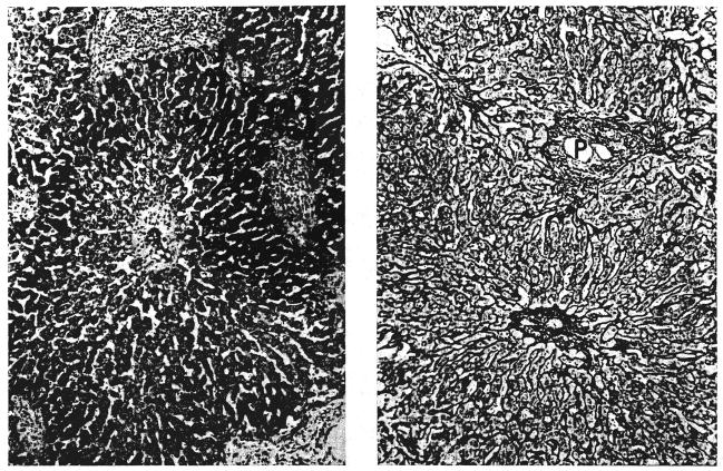
Two canine hepatic homegrafts: A, left, 120 days after transplantation (dog 5 group 1). A band of reticulin fibers links 2 central veins. Reticulin, × 65. B, right, 52 days after transplantation (dog 18 group 2). Centrizonal liver cell necrosis and hemorrhage is combined with cellular infiltration of the portal tracts (P). Compare with Figure 6A. Hematoxylin and eosin, × 65.

Operative angiograms on dog 20 of group 2. A, left, Contrast medium injected into the splenic vein passes through the animal’s own liver. B, right, Contrast material injected through a distal mesenteric vein tributary passes through the homografted liver. Note the relatively large size of the homograft compared to the animal’s own liver. This impression was confirmed at autopsy at which the homograft weighed 346 grams and the autologous liver, 262 grams.
Clinical course of dog 20 of group 2. The animal is the same one as shown in Figure 8.
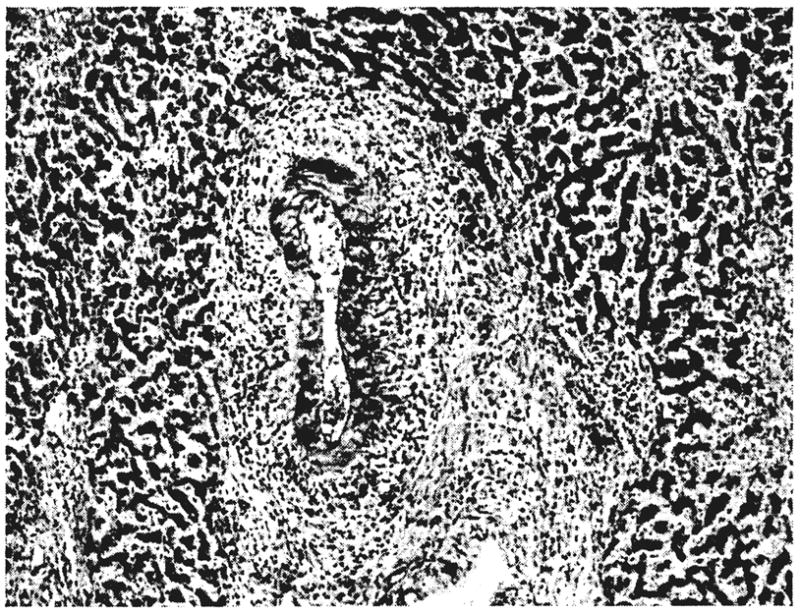
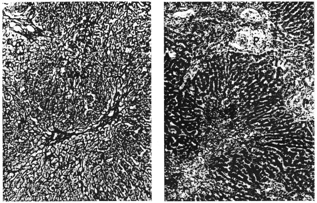
Homografted liver 50 days after transplantation (dog 19 group 2). In the portal tract a branch of the hepatic artery shows fibrinoid necrosis of its wall. The surrounding connective tissue is infiltrated with cells. At the right of the photomicrograph there is centrizonal necrosis of liver cells. Hematoxylin and eosin, × 50.
References
-
- Bodansky A. Notes on the determination of inorganic phosphate and serum phosphatase activity. J Biol Chem. 1937;120:167.
-
- Bras G, Jelliffe DB, Stuart KL. Venoocclusive disease of the liver with non-portal type of cirrhosis occurring in Jamaica. Arch Path Chic. 1954;57:285. - PubMed
-
- Goodrich EO, Welch HF, Nelson JA, Beecher TA, Welch GS. Homotransplantation of the canine liver. Surgery. 1956;39:244. - PubMed
-
- Malloy HT, Evelyn KA. The determination of bilirubin with the photoelectric colorimeter. J Biol Chem. 1937;119:481.
MeSH terms
Grants and funding
LinkOut - more resources
Full Text Sources
Other Literature Sources
Miscellaneous