. 2003 Oct;71(10):5488-97.
doi: 10.1128/IAI.71.10.5488-5497.2003.

Interleukin-18 facilitates the early antimicrobial host response to Escherichia coli peritonitis

Affiliations

Interleukin-18 facilitates the early antimicrobial host response to Escherichia coli peritonitis

Sebastiaan Weijer et al. Infect Immun. 2003 Oct.

Abstract

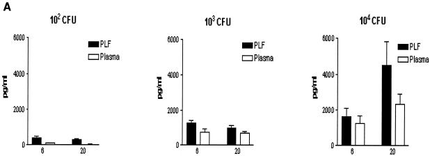
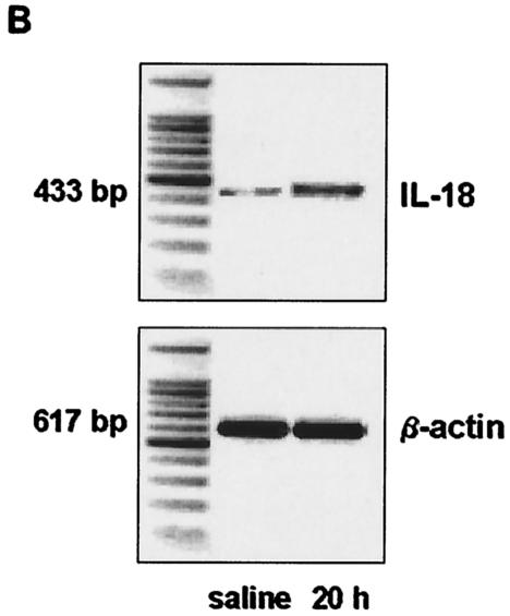
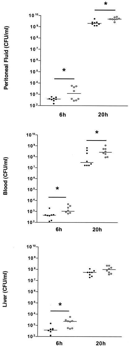
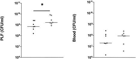
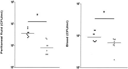
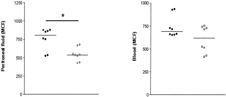
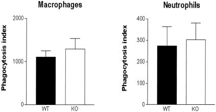
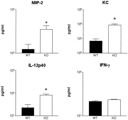
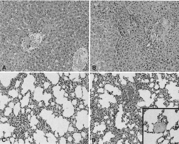
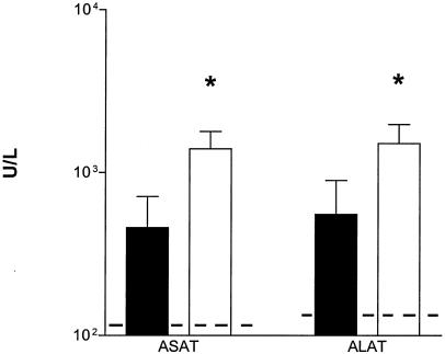
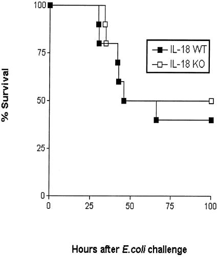
To determine the role of endogenous interleukin-18 (IL-18) during peritonitis, IL-18 gene-deficient (IL-18 KO) mice and wild-type mice were intraperitoneally (i.p.) infected with Escherichia coli, the most common causative agent found in septic peritonitis. Peritonitis was associated with a bacterial dose-dependent increase in IL-18 concentrations in peritoneal fluid and plasma. After infection, IL-18 KO mice had significantly more bacteria in the peritoneal lavage fluid and were more susceptible for progression to systemic infection at 6 and 20 h postinoculation than wild-type mice. The relative inability of IL-18 KO mice to clear E. coli from the abdominal cavity was not due to an intrinsic defect in the phagocytosing capacity of their peritoneal macrophages or neutrophils. IL-18 KO mice displayed an increased neutrophil influx into the peritoneal cavity, but these migratory neutrophils were less activate, as reflected by a reduced CD11b surface expression. These data suggest that endogenous IL-18 plays an important role in the early antibacterial host response during E. coli-induced peritonitis.


Figures

FIG. 1.
FIG. 1.
Enhanced IL-18 production during peritonitis. (A). IL-18 levels, measured by ELISA, in peritoneal lavage fluid (PLF) and plasma. WT mice were injected i.p. with 200 μl containing 102, 103, and 104 CFU of E. coli and sacrificed after 6 and 20 h. Results are expressed as mean and standard error of the mean for eight mice per group. (B). IL-18 mRNA and β-actin mRNA expression in the liver as determined by RT-PCR at 20 h after i.p. injection of sterile saline or 104 CFU of E. coli. Livers from three mice per group were pooled.
FIG. 1.
FIG. 1.
Enhanced IL-18 production during peritonitis. (A). IL-18 levels, measured by ELISA, in peritoneal lavage fluid (PLF) and plasma. WT mice were injected i.p. with 200 μl containing 102, 103, and 104 CFU of E. coli and sacrificed after 6 and 20 h. Results are expressed as mean and standard error of the mean for eight mice per group. (B). IL-18 mRNA and β-actin mRNA expression in the liver as determined by RT-PCR at 20 h after i.p. injection of sterile saline or 104 CFU of E. coli. Livers from three mice per group were pooled.
FIG. 2.
FIG. 2.
IL-18 KO mice demonstrate enhanced bacterial outgrowth. The E. coli cell count in peritoneal lavage fluid (top panel), blood (middle panel), and liver (bottom panel) in IL-18 KO and WT mice 6 and 20 h after i.p. administration of 104 CFU of E. coli is shown. Solid symbols represent WT mice; open symbols represent IL-18 KO mice. Horizontal lines represent medians. *, P < 0.05 for the difference between groups.
FIG. 3.
FIG. 3.
Anti-IL-18 treatment increases bacterial outgrowth. The E. coli cell count in peritoneal lavage fluid (left) and blood (right) in WT mice 20 h after i.p. administration of 104 CFU of E. coli is shown. At 1 h prior to infection, mice received either rabbit anti-IL-18 serum (200 μl) (open symbols) or normal rabbit serum (control) (solid symbols). Horizontal lines represent medians. *, P < 0.05 for the difference between groups.
FIG. 4.
FIG. 4.
Recombinant IL-18 inhibits bacterial outgrowth. The E. coli cell count in peritoneal lavage fluid (left) and blood (right) in WT mice 20 h after i.p. administration of 104 CFU of E. coli is shown. At 1 h prior to infection, mice received either 0.1 μg of recombinant mouse IL-18 (open symbols) or saline (control) (solid symbols). Horizontal lines represent medians. *, P < 0.05 for the difference between groups.
FIG. 5.
FIG. 5.
IL-18 KO mice have a decreased CD11b expression on peritoneal neutrophils. CD11b expression (mean channel fluorescence [MCF]) was determined by FACS analysis of neutrophils harvested from peritoneal fluid at 20 h postinfection, as described in Materials and Methods. Solid symbols represent WT mice; open symbols indicate IL-18 KO mice. Horizontal lines represent medians. *, P < 0.05 for the difference between groups.
FIG. 6.
FIG. 6.
Phagocytosis of E. coli by peritoneal macrophages and neutrophils of IL-18 KO mice is unchanged with respect to WT. Phagocytosis of FITC-labeled E. coli by macrophages and granulocytes harvested from peritoneal fluid from uninfected mice was determined by FACS analysis as described in Materials and Methods. Solid bars represent WT mice; open bars represent IL-18 KO mice. Results (mean and standard error) are expressed as the phagocytosis index, defined as the percentage of cells with internalized E. coli multiplied by the mean fluorescence intensity.
FIG. 7.
FIG. 7.
Chemokine and cytokine concentrations in peritoneal lavage fluid. MIP-2, KC, IL-12p40 and IFN-γ concentrations in peritoneal lavage fluid obtained 20 h postinfection are shown. Solid bars represent WT mice; open bars represent IL-18 KO mice. Data are the mean and standard error for eight mice per strain. *, P < 0.05 versus WT mice.
FIG. 8.
FIG. 8.
IL-18 KO mice demonstrate increased liver and lung injury. Representative views of the histological damage in the livers (A and B) and lungs (C and D) of WT mice (A and C) and IL-18 KO mice (B and D) at 20 h after infection are shown. Liver necrosis was more extended in IL-18 KO mice than in WT mice. The lungs of IL-18 KO mice were also more inflamed than those of WT mice. Numerous thrombi were observed (inset in panel D). The illustrations shown are representative of a total of eight mice per group. Hematoxylin and eosin staining. Magnification, ×20 (A to D) and ×40 (inset in panel D).
FIG. 9.
FIG. 9.
IL-18 KO mice demonstrate enhanced hepatocellular injury. Concentrations of ALT and AST in plasma (units/liter) at 20 h after i.p. injection of E. coli (104 CFU) are shown. Solid bars represent WT mice; open bars represent IL-18 KO mice. Data are the mean and standard error for eight mice per strain. Dotted lines represent the mean values obtained from normal plasma of mice that were injected i.p. with sterile saline (six mice). *, P < 0.05 versus WT mice.
FIG. 10.
FIG. 10.
IL-18 deficiency does not influence survival during murine peritonitis. Survival of WT and IL-18 KO mice infected i.p. with E. coli (104 CFU; n = 10 per group). No difference was found between the mortality rate in the two mouse strains.

Similar articles

Cited by

References

    1. Akira, S. 2000. The role of IL-18 in innate immunity. Curr. Opin. Immunol. 12:59-63. - PubMed
    1. Akira, S. 2001. Toll-like receptors and innate immunity. Adv. Immunol. 78:1-56. - PubMed
    1. Arndt, P. G., G. Fantuzzi, and E. Abraham. 2000. Expression of interleukin-18 in the lung after endotoxemia or hemorrhage-induced acute lung injury. Am. J. Respir. Cell Mol. Biol. 22:708-713. - PubMed
    1. Bohn, E., A. Sing, R. Zumbihl, C. Bielfeldt, H. Okamura, M. Kurimoto, J. Heesemann, and I. B. Autenrieth. 1998. IL-18 (IFN-gamma-inducing factor) regulates early cytokine production in, and promotes resolution of, bacterial infection in mice. J. Immunol. 160:299-307. - PubMed
    1. Bosscha, K., K. Reijnders, P. F. Hulstaert, A. Algra, and C. van der Werken. 1997. Prognostic scoring systems to predict outcome in peritonitis and intra-abdominal sepsis. Br. J. Surg. 84:1532-1534. - PubMed

Publication types

MeSH terms