. 2003 Oct;71(10):5921-39.
doi: 10.1128/IAI.71.10.5921-5939.2003.

Acid-adaptive genes of Helicobacter pylori

Affiliations

Acid-adaptive genes of Helicobacter pylori

Yi Wen et al. Infect Immun. 2003 Oct.

Abstract

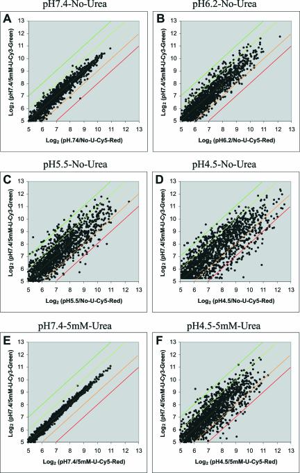
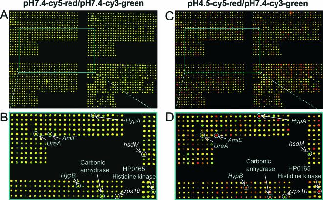
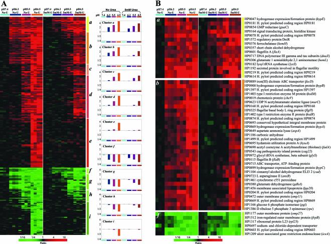
Helicobacter pylori is the only neutralophile that has been able to colonize the human stomach by using a variety of acid-adaptive mechanisms. One of the adaptive mechanisms is increased buffering due to expression of an acid-activated inner membrane urea channel, UreI, and a neutral pH-optimum intrabacterial urease. To delineate other possible adaptive mechanisms, changes in gene expression in response to acid exposure were examined using genomic microarrays of H. pylori exposed to different levels of external pH (7.4, 6.2, 5.5, and 4.5) for 30 min in the absence and presence of 5 mM urea. Gene expression was correlated with intrabacterial pH measured using 2',7'-bis-(2-carboxyethyl)-5-carboxyfluorescein and compared to that observed with exposure to 42 degrees C for 30 min. Microarrays containing the 1,534 open reading frames of H. pylori strain 26695 were hybridized with cDNAs from control (pH 7.4; labeled with Cy3) and acidic (labeled with Cy5) conditions. The intrabacterial pH was 8.1 at pH 7.4, fell to 5.3 at pH 4.5, and rose to 6.2 with urea. About 200 genes were up-regulated and approximately 100 genes were down-regulated at pH 4.5 in the absence of urea, and about half that number changed in the presence of urea. These genes included pH-homeostatic, transcriptional regulatory, motility, cell envelope, and pathogenicity genes. The up-regulation of some pH-homeostatic genes was confirmed by real-time PCR. There was little overlap with the genes induced by temperature stress. These results suggest that H. pylori has evolved multifaceted acid-adaptive mechanisms enabling it to colonize the stomach that may be novel targets for eliminating infection.


Figures

FIG. 1.
FIG. 1.
Scatter analysis of the general patterns of acidic responses of H. pylori genes revealed by transcriptional profiling. Scatter plots (log2-log2) of average normalized intensities representing Cy5-red channel versus Cy3-green channel are shown for experiments at pH 7.4 (n = 3) (A), 6.2 (n = 4) (B), 5.5 (n = 3) (C), and 4.5 (n = 4) (D) without urea (No-U), as well as pH 7.4 (n = 3) (E) and 4.5 (n = 4) (F) with 5 mM urea (U), compared with pH 7.4. Differential expression of a given gene is reflected by deviation from the yellow diagonal line. The orange diagonal defines ≥2-fold up-regulation, the red diagonal defines ≥4-fold up-regulation, light green defines ≥2-fold down-regulation, and dark green defines ≥4-fold down-regulation.
FIG. 2.
FIG. 2.
Representative hybridization with cDNA prepared from H. pylori strain ATCC 26695 incubated at pH 7.4 and 4.5. Microarrays containing 1,534 ORFs from H. pylori strain 26695 were hybridized with fluorescent Cy3-labeled H. pylori strain 26695 cDNA from control conditions (pH 7.4) and Cy5-labeled cDNAs from experimental conditions at pH 7.4 (A and B) and pH 4.5 with 5 mM urea (C and D). A close-up region (boxed in each array image) is also shown (B or D). Genes that are up- or down-regulated by low pH appear in the image as red or green spots, respectively. Genes that are expressed at similar levels in both samples appear as yellow spots. ureA (encoding the urease structural subunit) does not change, whereas amiE (encoding aliphatic amidase), HP1186 (encoding carbonic anhydrase), HypA and HypB (hydrogenase accessory proteins) genes, and HP0165 (encoding histidine kinase) show clear up-regulation, and the type II restriction enzyme gene, hsdM, and a ribosomal protein gene, rps10, are down-regulated.
FIG. 3.
FIG. 3.
Cluster analysis of gene expression patterns at different medium pHs with and without urea. (A) Hierarchical clustering was applied to expression data from a set of 278 genes measured across eight experimental pH conditions. The 278 genes were those (of 1,534 total) with transcript levels that varied by at least twofold relative to the control condition (pH 7.4) under conditions of pH 4.5 without urea. Data from 28 hybridizations were used: 3 for pH 7.4, 4 for pH 6.2, 3 for pH 5.5, and 4 for pH 4.5 without urea, as well as 3 for pH 7.4, 3 for pH 6.2, 4 for pH 5.5, and 4 for pH 4.5 with 5 mM urea. The color scale used to represent the expression ratios is shown on the bottom. The colored bars with labels (a to i) refer to the identified clusters of genes. A profile bar graph for each cluster in which the mean log ratio of that cluster in each experimental condition is plotted is shown on the right. (B) Enlargements of regions of the cluster diagram in panel A. The genes in up-regulated cluster a (top) and down-regulated cluster f (bottom), beginning at pH 6.2 and independent of the presence of urea, are shown. The genes up-regulated at pH 6.2 where the presence of urea results in up-regulation only when the medium pH is 4.5 are shown in cluster b (middle image). Gene names are shown corresponding to their positions in the cluster diagram.
FIG. 4.
FIG. 4.
Real-time PCR. (A to C) The cDNAs encoding periplasmic carbonic anhydrase (A), aliphatic amidase (B), and arginase (C) from typical experiments of the microarray analysis at pH 7.4 and 4.5 in the absence of urea were subjected to PCR amplification. The lines represent amplifications from cDNAs generated from the RNAs isolated at pH 7.4 and 4.5. (D) Similar amplification for the amidase cDNA resulting from temperature stress (42°C) compared to the cDNA generated from the RNA isolated from bacteria maintained at 37°C.

References

    1. Akada, J. K., M. Shirai, H. Takeuchi, M. Tsuda, and T. Nakazawa. 2000. Identification of the urease operon in Helicobacter pylori and its control by mRNA decay in response to pH. Mol. Microbiol. 36:1071-1084. - PubMed
    1. Allan, E., C. L. Clayton, A. McLaren, D. M. Wallace, and B. W. Wren. 2001. Characterization of the low-pH responses of Helicobacter pylori using genomic DNA arrays. Microbiology 147:2285-2292. - PubMed
    1. Ang, S., C. Z. Lee, K. Peck, M. Sindici, U. Matrubutham, M. A. Gleeson, and J. T. Wang. 2001. Acid-induced gene expression in Helicobacter pylori: study in genomic scale by microarray. Infect. Immun. 69:1679-1686. - PMC - PubMed
    1. Athmann, C., N. Zeng, T. Kang, E. A. Marcus, D. R. Scott, M. Rektorschek, A. Buhmann, K. Melchers, and G. Sachs. 2000. Local pH elevation mediated by the intrabacterial urease of Helicobacter pylori cocultured with gastric cells. J. Clin. Investig. 106:339-347. - PMC - PubMed
    1. Beier, D., and R. Frank. 2000. Molecular characterization of two-component systems of Helicobacter pylori. J. Bacteriol. 182:2068-2076. - PMC - PubMed

Publication types

Substances

LinkOut - more resources