. 2003 Oct;71(10):5940-50.
doi: 10.1128/IAI.71.10.5940-5950.2003.

An attenuated strain of the facultative intracellular bacterium Francisella tularensis can escape the phagosome of monocytic cells

Affiliations

An attenuated strain of the facultative intracellular bacterium Francisella tularensis can escape the phagosome of monocytic cells

Igor Golovliov et al. Infect Immun. 2003 Oct.

Abstract

The facultative intracellular bacterium Francisella tularensis is a highly virulent and contagious organism, and little is known about its intracellular survival mechanisms. We studied the intracellular localization of the attenuated human vaccine strain, F. tularensis LVS, in adherent mouse peritoneal cells, in mouse macrophage-like cell line J774A.1, and in human macrophage cell line THP-1. Confocal microscopy of infected J774A.1 cells indicated that during the first hour of infection the bacteria colocalized with the late endosomal-lysosomal glycoprotein LAMP-1, but within 3 h this colocalization decreased significantly from approximately 60% to 30%. Transmission electron microscopy revealed that >90% of bacteria were not enclosed by a phagosomal membrane after 2 h of infection, and some bacteria were in vacuoles that were only partially surrounded by a limiting membrane. Similar findings were obtained with all three host cell types. Immunoelectron microscopy performed with an F. tularensis LVS-specific polyclonal rabbit antiserum showed that the antiserum stained a thick, evenly distributed capsule-like material in bacteria grown in broth. In contrast, intracellular F. tularensis LVS cells were only marginally stained with this antiserum. Instead, most of the immunoreactive material was diffusely localized in the phagosomes or was associated with the phagosomal membrane. Our findings indicate that F. tularensis LVS is able to escape from the phagosomes of macrophages via a mechanism that may involve degradation of the phagosomal membrane.


Figures

FIG. 1.
FIG. 1.
Kinetics of colocalization of F. tularensis LVS-GFP or S. enterica GFP and the late endosomal-lysosomal glycoprotein LAMP-1. J774A.1 cells were infected with F. tularensis LVS or S. enterica at an MOI of 50 for 60 min, and extracellular bacteria were removed by washing (time zero). Infected cells were then fixed or incubated for another 1 or 3 h before fixation. They were then stained for LAMP-1 together with a Cy3-labeled secondary antibody. The data are means ± standard errors for at least 100 sections. Open bars, colocalization of S. enterica and LAMP-1; solid bars, colocalization of F. tularensis LVS and LAMP-1. The asterisk indicates a percentage which is significantly lower than that observed at time zero (P < 0.01).
FIG. 2.
FIG. 2.
Representative confocal microscopic images of localization of F. tularensis LVS-GFP or S. enterica GFP and the late endosomal-lysosomal marker LAMP-1 in J774A.1 cells. Macrophages were infected for 1 h with the bacteria and washed (time zero). Infected cells were then fixed or incubated for another 3 h before fixation. After fixation, they were stained for LAMP-1, and binding was visualized by using a Cy3-labeled secondary antibody.
FIG.3.
FIG.3.
Transmission electron micrographs of mouse J774A.1 macrophages infected with F. tularensis LVS at 2 h (A to D) and 18 h (E to H). (A) F. tularensis LVS localized in a phagosome. The arrowheads indicate a clearly visible phagosomal membrane. The arrow indicates the macrophage surface membrane. Magnification, ×39,000. (B) F. tularensis LVS localized in a phagosome with an incomplete membrane (arrowheads). Magnification, ×39,000. (C) F. tularensis LVS localized in the cytoplasm and surrounded by an electron-lucent space. The arrows indicate vesicular bodies in the space. Magnification, ×39,000. (D) F. tularensis LVS localized in the cytoplasm. Note the presence of numerous vesicular and membranous bodies (arrows) in a surrounding electron-translucent space. Magnification, ×39,000. (E) Numerous F. tularensis LVS cells (stars) in the cytoplasm. Magnification, ×26,000. (F) F. tularensis LVS cells localized in the cytoplasm. The arrow indicates a bacterium. Magnification, ×26,000. (G) F. tularensis LVS-infected macrophage with numerous bacteria in the cytoplasm. N, nucleus. Magnification, ×6,000. (H) F. tularensis LVS-infected macrophage with numerous bacteria in the cytoplasm, showing ultrastructural signs of apoptosis, cytoplasmic condensation, chromatin aggregation and marginalization, and cell surface blebbing. N, nucleus. Magnification, ×5,500. (I) Detail of panel G at a higher magnification (×24,000).
FIG. 4.
FIG. 4.
Transmission electron micrographs of mouse J774A.1 macrophages infected with F. tularensis LVS at 2 h. The macrophages contain single bacteria in the cytoplasm (enclosed in boxes). The insets show higher magnifications of the bacteria. The bacteria are separated from the cytosol only by a narrow electron-lucent space. (A) Magnification, ×12,000 (inset, ×27,000). (B) Magnification, ×10,000 (inset, ×28,000).
FIG. 5.
FIG. 5.
Transmission electron micrographs of mouse J774A.1 macrophages infected with S. enterica serovar Typhimurium at 2 h. (A) S. enterica (arrow) localized in a phagosome. Magnification, ×16,000. (B) Arrowheads indicate an intact phagosomal membrane. Magnification, ×43,000.
FIG. 6.
FIG. 6.
Transmission electron micrographs of adherent mouse PEC (A to C) and human THP-1 cells (D to F) infected with F. tularensis LVS for 2 h. (A) F. tularensis LVS localized in a phagosome. The arrowheads indicate a clearly visible phagosomal membrane. Magnification, ×30,000. (B) Two F. tularensis LVS cells partially surrounded by phagosomal membrane fragments (arrowheads) and electron-lucent space containing vesicular and membranous bodies (arrows). Magnification, ×32,000. (C) F. tularensis LVS (asterisk) in a perinuclear area, possibly escaping from the phagosome. Note the absence of a phagosomal membrane around the bacterium. Magnification, ×35,000. (D) F. tularensis LVS completely enclosed by the phagosomal membrane (arrowheads). Magnification, ×40,000. (E) The arrow indicates F. tularensis LVS localized in the cytoplasm and separated from the cytoplasm by an electron-translucent space. For comparison, an adjacent phagosome lacking bacteria contains a complete membrane (arrowheads). Magnification, ×40,000. (F) F. tularensis LVS localized in the cytoplasm (asterisk). Note the presence of vesicular and polymorphic bodies in the surrounding clear space (arrows). Magnification, ×41,000.
FIG. 7.
FIG. 7.
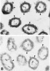
Immunoelectron microscopic localization of the anti-FT-reactive material in extracellular F. tularensis LVS. (A) The electron-dense peroxidase reaction product stains a thick surface layer (arrowheads) of extracellular F. tularensis LVS cells. (B) No bacterial surface staining was observed when anti-FT serum was replaced with normal rabbit serum (arrowheads). Magnification, ×32,000. The ultrathin sections were examined without any additional staining.
FIG. 8.
FIG. 8.
Immunoelectron microscopic localization of the anti-FT-reactive material in infected J774A.1 macrophages at 2 h. (A) F. tularensis LVS localized in the phagosome. The electron-dense reaction product partially stains the bacterial surface (arrows). (B) The reaction product stains some areas of the bacterial envelope (arrow) and amorphous contents of the phagosome (arrowheads). (C) F. tularensis LVS showing scarce clusters of surface deposits of the reaction product (arrow). Simultaneously the reaction product partially stains the phagosomal membrane (arrowheads). Note the triple structure of the bacterial envelope (open arrow). (D) The anti-FT-reactive bacterial material labels the phagosomal limiting membrane (arrowheads). (E) Anti-FT serum does not stain an F. tularensis LVS cell. Instead, it stains a phagosomal membrane (arrowheads) that is partially fragmented (thick arrow). The thin arrow indicates stained microvesicles. (F) F. tularensis LVS cell not stained by anti-FT serum localized in the cytoplasm. The cell is surrounded by electron-lucent space which contains labeled microvesicular and membranous inclusions (arrows) morphologically resembling those shown in Fig. 4C and D. (G) Control with labeling of bacteria in the absence of specific antiserum. Magnification, ×41,000. The ultrathin sections were examined without any additional staining.

References

    1. Alvarez-Dominguez, C., R. Roberts, and P. D. Stahl. 1997. Internalized Listeria monocytogenes modulates intracellular trafficking and delays maturation of the phagosome. J. Cell Sci. 110:731-743. - PubMed
    1. Amer, A. O., and M. S. Swanson. 2002. A phagosome of one's own: a microbial guide to life in the macrophage. Curr. Opin. Microbiol. 5:56-61. - PubMed
    1. Anthony, L. S., P. J. Morrissey, and F. E. Nano. 1992. Growth inhibition of Francisella tularensis live vaccine strain by IFN-gamma-activated macrophages is mediated by reactive nitrogen intermediates derived from l-arginine metabolism. J. Immunol. 148:1829-1834. - PubMed
    1. Anthony, L. S. D., R. D. Burke, and F. E. Nano. 1991. Growth of Francisella spp. in rodent macrophages. Infect. Immun. 59:3291-3296. - PMC - PubMed
    1. Camilli, A., H. Goldfine, and D. A. Portnoy. 1991. Listeria monocytogenes mutants lacking phosphatidylinositol-specific phospholipase C are avirulent. J. Exp. Med. 173:751-754. - PMC - PubMed

Publication types

MeSH terms

LinkOut - more resources