. 2003 Dec 1;553(Pt 2):545-59.
doi: 10.1113/jphysiol.2003.050419. Epub 2003 Sep 18.

Pacing of interstitial cells of Cajal in the murine gastric antrum: neurally mediated and direct stimulation

Affiliations

Pacing of interstitial cells of Cajal in the murine gastric antrum: neurally mediated and direct stimulation

Elizabeth A H Beckett et al. J Physiol. .

Abstract

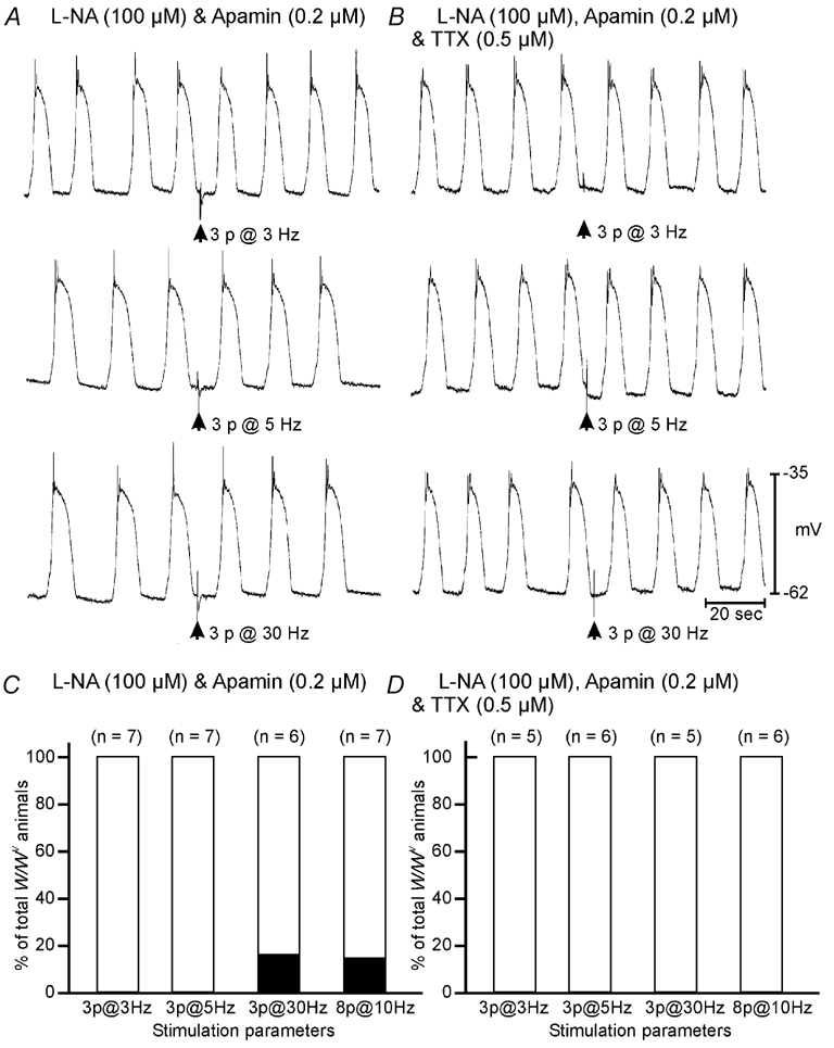
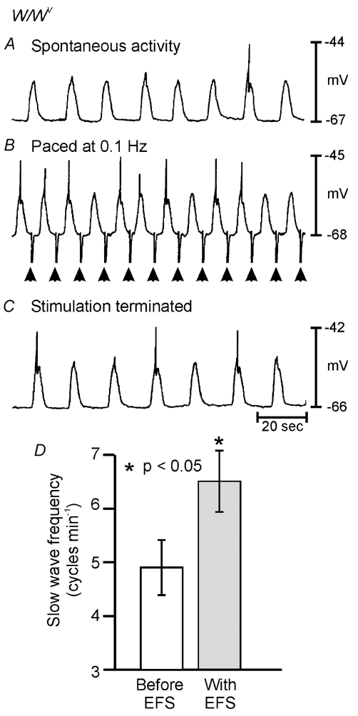
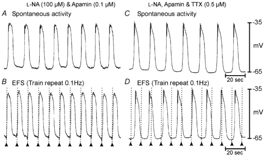
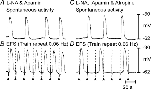
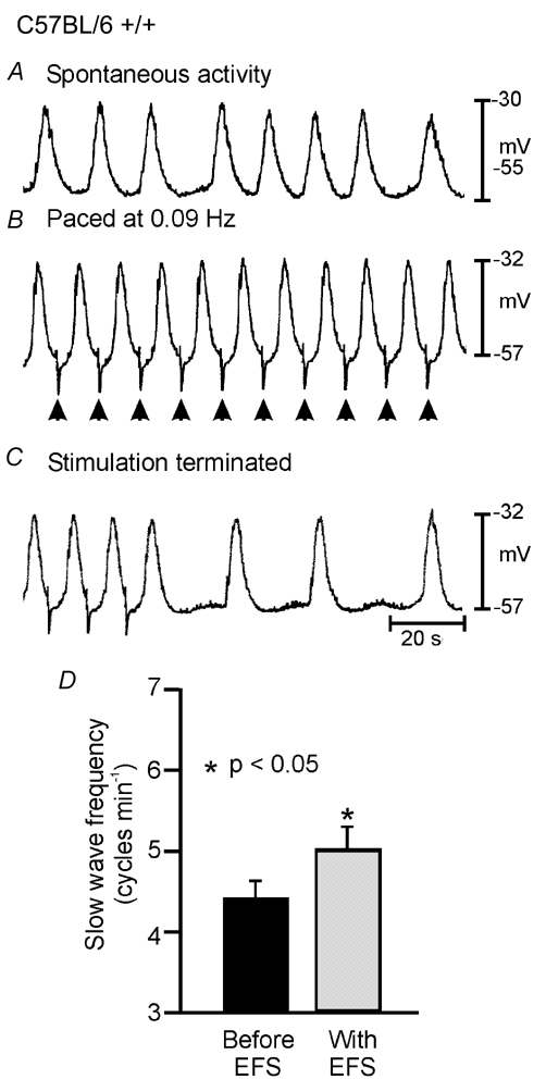
Phase advancement of electrical slow waves and regulation of pacemaker frequency was investigated in the circular muscle layer of the gastric antra of wild-type and W/W(V) mice. Slow waves in the murine antrum of wild-type animals had an intrinsic frequency of 4.4 cycles min(-1) and were phase advanced and entrained to a maximum of 6.3 cycles min(-1) using 0.1 ms pulses of electrical field stimulation (EFS) (three pulses delivered at 3-30 Hz). Pacing of slow waves was blocked by tetrodotoxin (TTX) and atropine, suggesting phase advancement was mediated via intrinsic cholinergic nerves. Phase advancement and entrainment of slow waves via this mechanism was absent in W/W(V) mutants which lack intramuscular interstitial cells of Cajal (ICC-IM). These data suggest that neural regulation of slow wave frequency and regulation of smooth muscle responses to slow waves are mediated via nerve-ICC-IM interactions. With longer stimulation parameters (1.0-2.0 ms), EFS phase advanced and entrained slow waves in wild-type and W/W(V) animals. Pacing with 1-2 ms pulses was not inhibited by TTX or atropine. These data suggest that stimulation with longer pulse duration is capable of directly activating the pacemaker mechanism in ICC-MY networks. In summary, intrinsic excitatory neurons can phase advance and increase the frequency of antral slow waves. This form of regulation is mediated via ICC-IM. Longer pulse stimulation can directly activate ICC-MY in the absence of ICC-IM.


Figures

Figure 1
Figure 1. Kit-like immunohistochemistry revealed the distribution of specific classes of ICC in gastric antrum of BALB/c, C57BL/6 +/+ and W/WV mice
The density of kit-immunopositive interstitial cells of Cajal (ICC) at the level of the myenteric plexus (ICC-MY) was greatest in the region of the greater curvature in BALB/c(A), C57BL/6+/+ (C) and W/WV mice (E), as previously demonstrated (see Ordog et al. 2002; Hirst et al. 2002a). Spindle-shaped intramuscular ICC (ICC-IM) were distributed throughout the circular muscle layer and were in equal densities in both BALB/c and C57BL/6 wild-type mice (B and D). In contrast, ICC-IM were absent within the circular muscle layer of W/WV animals (F).
Figure 2
Figure 2. ICC-IM and intrinsic cholinergic motor nerves are closely apposed within the circular muscle layer of the gastric antrum
In wild-type animals, immunohistochemical labelling for Kit (Kit-Li; A; red) and vesicular acetylcholine transporter (vAChT-Li; B; green) revealed that nerve bundles containing vAChT-positive fibres were closely associated with ICC-IM (red). (C, overlay of micrographs A and B). A higher magnification of the close apposition between vAChT-Li nerves and ICC-IM is shown in D. Despite the absence of Kit-immunopositive ICC-IM in the circular muscle layer of W/WV mutants (E), the distribution of vAChT-Li enteric motor nerves was similar to that of wild-type strains (F). (G, overlay of micrographs E and F).
Figure 7
Figure 7. Short duration (0.1 ms) pulses of EFS fail to elicit premature slow wave events in W/WV antrum muscles
A, in the presence of l-NA (100 µm) and apamin (0.2 µm) slow waves from a W/WV antrum could not be phase advanced using 0.1 ms duration pulses of EFS delivered at frequencies of between 3 and 30 Hz. B, TTX did not alter the response to EFS. C, summarized data from all W/WV animals shows that failure of phase advancement was consistent, with only one W/WV antrum tissue showing statistical phase advancement following high frequency stimulation (3 pulses at 30 Hz and 8 pulses at 10 Hz). D, in the presence of TTX, slow waves could not be phase advanced at any frequency in W/WV animals. Scale bars at the bottom of B apply to all panels.
Figure 10
Figure 10. Slow waves in W/WV antrum muscles can be phase advanced using longer duration pulses of EFS (1–2 ms pulse duration)
A, in the presence of l-NA (100 µm) and atropine (1 µm) single 1.0 ms duration pulses of EFS (delivered at arrow) evoked fast inhibitory junction potentials and premature slow wave events. B, individual slow wave cycle periods (SWCPs) varied from cycle to cycle around a mean value of 19 s. The SWCP between the slow wave immediately preceding EFS and that following stimulation was 14 s significantly shorter than the mean SWCP (depicted by the encircled filled circle in B). C and D, TTX blocked the large inhibitory junction potential evoked by EFS but did not inhibit the phase advancement of slow waves following EFS. E, summary of data from 10 W/WV animals, analysed as described for Fig. 8. In 9 out of 10 W/WV animals the SWCP between the slow wave immediately preceding EFS and that following stimulation fell below the 95 % confidence limits of the mean SWCP, indicating that phase advancement had occurred (E).
Figure 8
Figure 8. Phase advancement of slow waves using long duration pulses of EFS (1–2 ms pulse duration) does not require intrinsic nerve activation
A, in the presence of l-NA (100 µm) and atropine (1 µm) single 1.0 ms duration pulses of EFS (delivered at arrow) evoked premature slow wave events. B, slow wave cycle periods (SWCPs) between consecutive slow waves varied from cycle to cycle around a mean value of 16 s. The SWCP between the slow wave immediately preceding EFS and that following stimulation was significantly shorter than the mean SWCP by 12 s (depicted by the encircled filled circle in B). C and D, slow wave advancement in the presence of l-NA, atropine and TTX (0.3 µm). The remaining voltage transient is a stimulus artifact. E, summary of data from 10 wild-type animals. For each animal, the SWCP of five spontaneous slow waves prior to electrical field stimulation were measured to determine the 95 % confidence limits of the mean SWCP. The mean SWCP was expressed as 100 % and is represented by the dashed line. Each of the 10 vertical lines represents the 95 % confidence intervals of the mean SWCP for an individual animal. The SWCP between the slow wave immediately preceding EFS and that following stimulation was expressed as a percentage of the mean SWCP (filled diamonds). If this value fell below the 95 % confidence limits of the mean SWCP it was considered that phase advancement had occurred. In all 10 wild-type animals single 1.0–2.0 ms pulses of EFS produced phase advancement of the next slow wave in the presence of l-NA, atropine and TTX (E).
Figure 11
Figure 11. Entrainment of W/WV antral slow waves using single 1.0–2.0 ms pulses of EFS
Spontaneous slow waves occurring at a rate of 4 cycles min−1 (0.067 Hz; A) were entrained at a frequency of 6 cycles min−1 (0.1 Hz; B) by single pulses of EFS (1.0 ms; supra-optimal voltage; delivered at arrows). C, slow wave discharge returned to the intrinsic frequency of 0.067 Hz soon after termination of EFS. D, summary of data from W/WV entrainment experiments. The mean intrinsic frequency of slow wave discharge from W/WV animals was 4.9 ± 0.5 cycles min−1 (open bar) and was increased to 6.5 ± 0.6 cycles min−1 using 1.0–2.0 ms EFS (grey bar; n = 9). Entrainment experiments using 1.0–2.0 ms pulses of EFS were performed in the presence of l-NA (100 µm), atropine (1 µm) and TTX (0.3 µm).
Figure 3
Figure 3. Slow wave events can be phase advanced by short duration (0.1 ms) pulses of electrical field stimulation (EFS) in wild-type antrum muscles
A, in the presence of Nω-nitro-l-arginine (l-NA) (100 µm) and apamin (0.2 µm) slow wave events were prematurely evoked by trains of short duration pulses (0.1 ms; 3–8 pulses at frequencies of 3–30 Hz; delivered at arrows). B, tetrodotoxin (0.5 µm) abolished the phase advancement of slow waves. C, summarized data from wild-type animals reveals that the advancement of slow wave events in the presence of l-NA and apamin was frequency dependent. As stimulation frequency was increased the percentage of animals in which phase advancement occurred increased (i.e. 54 % of wild-type animals showed phase advancement with 3 pulses at 3 Hz, whereas 80 % showed phase advancement with 3 pulses at 30 Hz (C)). In the presence of TTX (0.5 µm), slow waves could not be phase advanced at any frequency except in one tissue at a stimulus parameter of 8 pulses at 10 Hz (D). Scale bars at the bottom of B apply to all panels.
Figure 4
Figure 4. Slow waves can be paced by short duration (0.1 ms) pulses of EFS in wild-type antrum muscles
A, in the presence of l-NA (100 µm) and apamin (0.2 µm) slow waves occurred spontaneously at a frequency of 0.07 Hz (4.4 cycles min−1). B, trains of short duration pulses (0.1 ms; 3 pulses at 5 Hz; supra-optimal voltage) delivered at a frequency of 0.1 Hz entrained slow waves at a rate of 6 cycles min−1. Application of tetrodotoxin (TTX; 0.5 µm) did not alter spontaneous slow wave frequency (C) but inhibited electrical entrainment of slow waves (D). Time scale bars are indicated in panels C and D.
Figure 5
Figure 5. Blockade of muscarinic receptors prevents gastric antrum entrainment
Spontaneous slow waves recorded from the greater curvature of the terminal antrum (A) could be paced up to twice the intrinsic rate using trains of EFS (0.1 ms pulse duration; 3 pulses at 5 Hz; supra-optimal voltage) (B). Atropine (1 µm) did not significantly alter spontaneous slow wave frequency (C) but prevented slow wave entrainment at higher frequencies (D). Timescale bar shown in D is the same for all panels.
Figure 6
Figure 6. The slow wave cycle period was more variable in W/WV animals than their wild-type siblings
A, slow waves recorded from the terminal antrum of a C57BL/6 wild-type control animal. The straight line in B represents the mean slow wave cycle period (SWCP) calculated from 15 consecutive slow waves. From cycle to cycle, individual SWCPs (represented by filled circles) varied around a mean of 16 s (B). C, discharge of slow waves from the terminal antrum of W/WV animals was generally more irregular. D, demonstration of the variance of individual SWCPs (filled circles) from the mean SWCP value (continuous line). E, examination of W/WV slow waves on an expanded time scale reveals two components to the slow waves as previously described in murine antral muscles (e.g. Dickens et al. 2001); arrows denote inflection in upstroke potential separating the 1st and 2nd components.
Figure 9
Figure 9. Longer duration pulses of EFS (1.0–2.0 ms) produced entrainment of slow waves in wild-type antrum muscles
In the presence of l-NA (100 µm), atropine (1 µm) and TTX (0.5 µm) spontaneous slow waves which discharged at an intrinsic rate of 0.067 Hz (4 cycles min−1; A) could be entrained at 0.09 Hz (5.4 cycles min−1; B) by single 1.0 ms pulses of EFS (supra-optimal voltage). C, the slow wave frequency returned to its intrinsic rate within 1 min after EFS was terminated. D, summary of data from wild-type entrainment experiments. The mean slow wave frequency for wild-type animals was 4.4 ± 0.2 cycles min−1 (black bar; n = 12) and this was increased using single pulse EFS (1.0–2.0 ms pulse duration) to a mean value of 5.0 ± 0.3 cycles min−1 (grey bar).

References

    1. Banks BE, Brown C, Burgess GM, Burnstock G, Claret M, Cocks TM, Jenkinson DH. Apamin blocks certain neurotransmitter-induced increases in potassium permeability. Nature. 1979;282:415–417. - PubMed
    1. Beckett EA, Horiguchi K, Khoyi M, Sanders KM, Ward SM. Loss of enteric motor neurotransmission in the gastric fundus of Sl/Sl(d) mice. J Physiol. 2002;543:871–887. - PMC - PubMed
    1. Bortolotti M. The ‘electrical way’ to cure gastroparesis. Am J Gastroenterol. 2002;97:1874–1883. - PubMed
    1. Burns AJ, Lomax AE, Torihashi S, Sanders KM, Ward SM. Interstitial cells of Cajal mediate inhibitory neurotransmission in the stomach. Proc Natl Acad Sci U S A. 1996;93:12008–12013. - PMC - PubMed
    1. Daniel EE, Posey-Daniel V. Neuromuscular structures in opossum esophagus: role of interstitial cells of Cajal. Am J Physiol. 1984;246:G305–315. - PubMed

Publication types

MeSH terms

LinkOut - more resources